WO2010114029A1 - Method for detecting antibody against sith-1 in biological sample - Google Patents
Method for detecting antibody against sith-1 in biological sample Download PDFInfo
- Publication number
- WO2010114029A1 WO2010114029A1 PCT/JP2010/055884 JP2010055884W WO2010114029A1 WO 2010114029 A1 WO2010114029 A1 WO 2010114029A1 JP 2010055884 W JP2010055884 W JP 2010055884W WO 2010114029 A1 WO2010114029 A1 WO 2010114029A1
- Authority
- WO
- WIPO (PCT)
- Prior art keywords
- protein
- sith
- biotin
- carrier
- binding
- Prior art date
Links
Images


Classifications
-
- C—CHEMISTRY; METALLURGY
- C07—ORGANIC CHEMISTRY
- C07K—PEPTIDES
- C07K14/00—Peptides having more than 20 amino acids; Gastrins; Somatostatins; Melanotropins; Derivatives thereof
- C07K14/005—Peptides having more than 20 amino acids; Gastrins; Somatostatins; Melanotropins; Derivatives thereof from viruses
-
- G—PHYSICS
- G01—MEASURING; TESTING
- G01N—INVESTIGATING OR ANALYSING MATERIALS BY DETERMINING THEIR CHEMICAL OR PHYSICAL PROPERTIES
- G01N33/00—Investigating or analysing materials by specific methods not covered by groups G01N1/00 - G01N31/00
- G01N33/48—Biological material, e.g. blood, urine; Haemocytometers
- G01N33/50—Chemical analysis of biological material, e.g. blood, urine; Testing involving biospecific ligand binding methods; Immunological testing
- G01N33/53—Immunoassay; Biospecific binding assay; Materials therefor
- G01N33/543—Immunoassay; Biospecific binding assay; Materials therefor with an insoluble carrier for immobilising immunochemicals
-
- G—PHYSICS
- G01—MEASURING; TESTING
- G01N—INVESTIGATING OR ANALYSING MATERIALS BY DETERMINING THEIR CHEMICAL OR PHYSICAL PROPERTIES
- G01N33/00—Investigating or analysing materials by specific methods not covered by groups G01N1/00 - G01N31/00
- G01N33/48—Biological material, e.g. blood, urine; Haemocytometers
- G01N33/50—Chemical analysis of biological material, e.g. blood, urine; Testing involving biospecific ligand binding methods; Immunological testing
- G01N33/53—Immunoassay; Biospecific binding assay; Materials therefor
- G01N33/569—Immunoassay; Biospecific binding assay; Materials therefor for microorganisms, e.g. protozoa, bacteria, viruses
- G01N33/56983—Viruses
- G01N33/56994—Herpetoviridae, e.g. cytomegalovirus, Epstein-Barr virus
-
- G—PHYSICS
- G01—MEASURING; TESTING
- G01N—INVESTIGATING OR ANALYSING MATERIALS BY DETERMINING THEIR CHEMICAL OR PHYSICAL PROPERTIES
- G01N33/00—Investigating or analysing materials by specific methods not covered by groups G01N1/00 - G01N31/00
- G01N33/48—Biological material, e.g. blood, urine; Haemocytometers
- G01N33/50—Chemical analysis of biological material, e.g. blood, urine; Testing involving biospecific ligand binding methods; Immunological testing
- G01N33/68—Chemical analysis of biological material, e.g. blood, urine; Testing involving biospecific ligand binding methods; Immunological testing involving proteins, peptides or amino acids
- G01N33/6854—Immunoglobulins
-
- C—CHEMISTRY; METALLURGY
- C07—ORGANIC CHEMISTRY
- C07K—PEPTIDES
- C07K2319/00—Fusion polypeptide
- C07K2319/70—Fusion polypeptide containing domain for protein-protein interaction
-
- C—CHEMISTRY; METALLURGY
- C12—BIOCHEMISTRY; BEER; SPIRITS; WINE; VINEGAR; MICROBIOLOGY; ENZYMOLOGY; MUTATION OR GENETIC ENGINEERING
- C12N—MICROORGANISMS OR ENZYMES; COMPOSITIONS THEREOF; PROPAGATING, PRESERVING, OR MAINTAINING MICROORGANISMS; MUTATION OR GENETIC ENGINEERING; CULTURE MEDIA
- C12N2710/00—MICROORGANISMS OR ENZYMES; COMPOSITIONS THEREOF; PROPAGATING, PRESERVING, OR MAINTAINING MICROORGANISMS; MUTATION OR GENETIC ENGINEERING; CULTURE MEDIA dsDNA viruses
- C12N2710/00011—Details
- C12N2710/16011—Herpesviridae
- C12N2710/16511—Roseolovirus, e.g. human herpesvirus 6, 7
- C12N2710/16522—New viral proteins or individual genes, new structural or functional aspects of known viral proteins or genes
-
- G—PHYSICS
- G01—MEASURING; TESTING
- G01N—INVESTIGATING OR ANALYSING MATERIALS BY DETERMINING THEIR CHEMICAL OR PHYSICAL PROPERTIES
- G01N2333/00—Assays involving biological materials from specific organisms or of a specific nature
- G01N2333/005—Assays involving biological materials from specific organisms or of a specific nature from viruses
- G01N2333/01—DNA viruses
- G01N2333/03—Herpetoviridae, e.g. pseudorabies virus
-
- G—PHYSICS
- G01—MEASURING; TESTING
- G01N—INVESTIGATING OR ANALYSING MATERIALS BY DETERMINING THEIR CHEMICAL OR PHYSICAL PROPERTIES
- G01N2469/00—Immunoassays for the detection of microorganisms
- G01N2469/20—Detection of antibodies in sample from host which are directed against antigens from microorganisms
Definitions
- the present invention qualifies antibodies of small protein encoded by the intermediate by the transcript of HHV-6 (SITH-1) encoded by a latent intermediate transcript of HHV-6 in a biological sample. And / or a method for quantitative detection. According to the method of the present invention, it is possible to detect an antibody of SITH-1 protein which is present in a trace amount in a biological sample and is usually difficult to detect.
- herpesviridae virus a double-stranded linear DNA having a molecular weight of 80 to 150 ⁇ 10 6 daltons is contained in an icosahedral capsid of about 100 nm in diameter consisting of 162 capsomeres around the core protein.
- the envelope surrounds the entire virus and has a size of about 150 to 200 nm.
- Herpesviruses are found in almost all mammals and amphibians, and in particular, the herpesviridae viruses that are hosted by humans are called human herpesviruses (HHV). HHV is classified into ⁇ (herpes simplex virus, varicella-zoster virus, etc.), ⁇ (cytomegalovirus, etc.), and ⁇ (EB virus, etc.) subfamily, respectively.
- herpes viruses are characterized by a “latent infection”. “Latent infection” refers to an infectious state in which a virus does not produce infectious virus particles in a host cell and continues to survive. In this latent infection, the virus gene and the presence of the virus gene are assisted. The gene product is retained in the host cell. It is known that herpesviruses showing latent infections are caused by the host being restarted for some reason (eg, aging, poor physical condition (including fatigue)), and the production of virus particles is resumed and a large amount of virus is replicated (re- activation).
- latent infection refers to an infectious state in which a virus does not produce infectious virus particles in a host cell and continues to survive. In this latent infection, the virus gene and the presence of the virus gene are assisted. The gene product is retained in the host cell. It is known that herpesviruses showing latent infections are caused by the host being restarted for some reason (eg, aging, poor physical condition (including fatigue)), and the production of virus particles is resumed and
- herpes virus will continue to infect if there is no abnormality in the host, but once the host's body is modulated and a host crisis is detected, it is reactivated to look for another healthy host
- herpes viruses In order to study the ecology of such herpes viruses, it is essential to understand the latent infection and reactivation of viruses.
- the herpesviruses only the EB virus belonging to ⁇ -herpesvirus has much knowledge about latent infection, and there are many unclear points regarding the others.
- Non-Patent Document 1 discloses that HHV-6 is latently infected in macrophages having a relatively high degree of differentiation in peripheral blood, and the site of latent infection of HHV-6 in the host is clarified.
- Non-Patent Document 2 describes that HHV-6 moves into the brain at a very high rate at the time of initial infection, resulting in persistent infection and latent infection.
- Non-Patent Document 3 discloses a gene (latent infection gene) that is expressed during latent infection of HHV-6, and suggests that the gene has a function of controlling latent infection and reactivation of the virus. Yes.
- Non-Patent Document 4 there is an “intermediate stage” in which the HHV-6 latent infection state is relatively stable and gene expression is active, and is encoded by the latent infection gene and this gene. It has been shown that proteins (latent infectious gene proteins) are expressed in large amounts. Furthermore, Non-Patent Document 5 describes that an antibody against a latent infectious gene protein whose expression is enhanced at an intermediate stage exists in the serum of patients with chronic fatigue syndrome.
- SITH-1 BY THE INTERMEDIATE TRANSCRIPP OF HHV-6 (SITH) -1 was identified.
- SITH-1 protein has a function of increasing intracellular calcium concentration
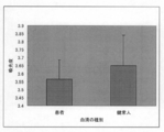
- SITH-1 The inventors found a new fact that antibodies against proteins are significantly detected in patients with mood disorders but rarely detected in healthy individuals, and filed a patent application (PCT / JP2008 / 67300).
- the serum antibody titer against the SITH-1 protein was measured by a conventional fluorescent antibody method using 293T cells expressing the SITH-1 protein as an antigen.
- the present inventors have continued further research, and the fluorescent antibody method requires the creation of SITH-1-expressing mammalian cells and the preparation of preparations, which requires a lot of labor.
- human serum against SITH-1 protein It has been found that the antibody titer is low, technical skill is required for the detection work, and that the SITH-1 protein is unstable and requires careful handling.
- the fluorescent antibody method is troublesome because fluorescence is visually determined under a microscope after reaction with a serum sample, and is unsuitable for simultaneous processing of many samples. Furthermore, non-specific binding becomes a problem for some serum samples, and measurement is often difficult.
- the present invention provides a method for simply, qualitatively and / or quantitatively detecting an antibody against SITH-1 protein in a biological sample.
- the purpose is to provide.
- the present invention provides a method for easily and qualitatively and / or quantitatively detecting and measuring an antibody against SITH-1 protein in a biological sample while suppressing a background signal. With the goal.
- the present inventors have found that a system in which SITH-1 protein is bound to a carrier is effective for antibody detection, and have come up with the present invention.
- the present invention provides a more sensitive method for detecting a SITH-1 protein antibody with reduced non-specific binding in a system in which a SITH-1 protein is immobilized on a carrier. .
- the present invention includes the following aspects.
- a method for detecting an antibody of Small protein encoded by the intermediate stage Transscript of HHV-6 (SITH-1) encoded by a latently infected intermediate transcript of HHV-6 in a biological sample 1) Prepare SITH-1 protein; 2) Binding the SITH-1 protein prepared in step 1) to a carrier; 3) The method comprising contacting a biological sample with the carrier bound with the SITH-1 protein prepared in step 2), and detecting the SITH-1 protein antibody.
- SITH-1 protein is selected from the following group: (A) a protein having the amino acid sequence of SEQ ID NO: 1; (B) a protein having an amino acid sequence including deletion, substitution, insertion and / or addition of one or more amino acids in the amino acid sequence of SEQ ID NO: 1 and having an activity of increasing intracellular calcium concentration; (C) a protein having an amino acid sequence having at least 80% identity to the amino acid sequence of SEQ ID NO: 1 and having an activity of increasing intracellular calcium concentration; (D) a protein having an amino acid sequence encoded by a nucleic acid consisting of the base sequence of SEQ ID NO: 2; (E) having an amino acid sequence encoded by a nucleic acid consisting of a base sequence containing a deletion, substitution, insertion and / or addition of one or more nucleotides in the base sequence of SEQ ID NO: 2 and having an intracellular calcium concentration A protein with increasing activity; (F) a protein having an amino acid sequence encoded by
- step 3) of embodiment 1 in the carrier bound with the SITH-1 protein prepared in step 2), (A) a biological sample, and (b) a cell disruption extract prepared from the same type of cells as the host cell used to express the SITH-1 protein in step 1) is mixed and added; Detection method according to aspect 1 or 2
- step 3) of embodiment 1 in the carrier bound with the SITH-1 protein prepared in step 2), (A) a biological sample and (bi) a host cell used to express the SITH-1 protein, biotinylated SITH-1 protein, and / or biotin-binding protein of step 1) or 2) Cell disruption extract prepared from the same type of cells and biotin-binding protein, or (b-ii) SITH-1 protein, biotinylated SITH-1 protein and / or biotin-binding protein of step 1) or 2) Cell disruption extract prepared from cells expressing biotin-binding protein by genetic engineering technology is added to cells of the same type as the host cell used for expression; Detection method according to aspect 4
- a carrier for detecting an antibody of Small protein encoded by the intermediate stage Transscript of HHV-6 (SITH-1) encoded by a latent intermediate transcript of HHV-6 in a biological sample A carrier to which the SITH-1 protein is bound.
- a kit for detecting an antibody of Small protein encoded by the intermediate stage Transscript of HHV-6 (SITH-1) encoded in a latent sample of latent infection of HHV-6 in a biological sample There, A) a carrier to which the SITH-1 protein is bound; and B) A kit comprising an agent for diluting a biological sample, comprising a cell disruption extract prepared from cells of the same type as the host cell used to express the SITH-1 protein of A).
- the carrier of A) is a carrier in which the SITH-1 protein is bound using a biotin-biotin-binding protein-protein bond
- the agent of B) i) A cell disruption extract prepared from cells of the same type as the host cell used to express the SITH-1 protein, biotinylated SITH-1 protein, and / or biotin-binding protein of A), and a biotin-binding protein Or ii) A biotin-binding protein is expressed by genetic engineering technology in the same type of host cell used to express the SITH-1 protein, biotinylated SITH-1 protein, and / or biotin-binding protein of A) An agent for diluting a biological sample, including a cell disruption extract prepared from the treated cells, The kit according to aspect 11.
- a kit for detecting an antibody of Small protein encoded by the intermediate stage Transscript of HHV-6 (SITH-1) encoded in a latent sample of latent infection of HHV-6 in a biological sample There, A) the SITH-1 protein; B) a carrier for immobilizing the SITH-1 protein of A); and C) a cell disruption extract prepared from cells of the same type as the host cell used to express the SITH-1 protein of A).
- A) SITH-1 protein is biotinylated, The carrier of B) is bound directly or indirectly to the biotin-binding protein, C) i) A cell disruption extract prepared from cells of the same type as the host cell used to express the SITH-1 protein, biotinylated SITH-1 protein, and / or biotin-binding protein of A) or B), and biotin Genetic engineering techniques to bind proteins, or ii) cells of the same type as the host cell used to express the SITH-1 protein, biotinylated SITH-1 protein, and / or biotin binding protein of A) or B) An agent for diluting a biological sample containing a cell disruption extract prepared from cells expressing a biotin-binding protein.
- detection of SITH-1 protein antibody in a biological sample can suppress background signal and stably perform detection with higher sensitivity. According to the method of the present invention, it is possible to detect and measure a SITH-1 protein antibody which is present in a trace amount in a biological sample and is difficult to detect by a usual method.
- FIG. 1 shows the expression of a BioEase tag (biotinylated tag) fused SITH-1 protein. Specifically, FIG. 1A shows the result of Western blotting for detection of SITH-1 protein, and FIG. 1B shows the result of activity staining for detection of biotinylated protein.
- E. coli BL21 (DE3) expressing the BioEase tag-fused SITH-1 protein was sonicated, and the resulting E. coli crude extract fraction was subjected to SDS-PAGE (15% acrylamide gel) to give 20 ⁇ g total protein / lane. And then transferred to a PVDF membrane.
- FIG. 1A reacted with anti-SITH-1 antibody (diluted 1/1000), reacted with alkaline phosphatase (AP) -labeled anti-rabbit IgG antibody (diluted 1/1000), and then stained with AP.
- FIG. 1B is a stained image after a streptavidin-horseradish peroxidase (HRP) (1/1000 dilution) reaction.
- HRP streptavidin-horseradish peroxidase
- Control indicates an extract sample derived from Escherichia coli having only an expression vector.
- the position of the BioEase tag-fused SITH-1 protein is indicated by an arrow.
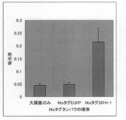
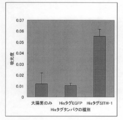
- FIG. 2 is a graph showing the effect of various serum dilutions on nonspecific binding. Serum diluted with PBS (white squares (dashed line)), diluted with PBS plus purified tamavidin 2 (TM2), white square (solid lines), diluted with E. coli disruption extract containing only expression vector The asterisk (broken line) is the asterisk (broken line), the asterisk (solid line) is diluted with the E.
- FIG. 3 is a graph showing the S / N ratio for each dilution ratio of the anti-SITH-1 antibody based on the results shown in FIG.
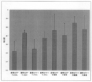
- FIG. 4 shows the results of measurement of anti-SITH-1 antibody titer in human serum using TM2 plates bound with biotinylated SITH-1.
- FIG. 5 shows the results of measurement of anti-SITH-1 antibody titer in human serum using TM2 plates bound with biotinylated SITH-1.
- Human serum diluted with an Escherichia coli crude extract was brought into contact with the TM2 plate.
- FIG. 6 shows the results of measurement of anti-SITH-1 antibody titer in human serum using TM2 plates bound with biotinylated SITH-1.
- FIG. 7 shows the results of anti-SITH-1 antibody titer measurement in anti-SITH-1 rabbit serum using a nickel plate to which a His tag-fused SITH-1 protein was bound.
- Anti-SITH-1 rabbit serum was diluted 100-fold with PBS (-) containing 0.2% BSA.
- PBS PBS
- E. coli not expressing anything and His-tagged EGFP were used.
- FIG. 8 shows the results of anti-SITH-1 antibody titer measurement in anti-SITH-1 rabbit serum using a nickel plate to which a His tag-fused SITH-1 protein was bound.
- Anti-SITH-1 rabbit serum was diluted 500-fold with PBS (-) containing 0.2% BSA.
- FIG. 9 shows the results of anti-SITH-1 antibody titer measurement in human serum when using a nickel plate to which a His tag-fused SITH-1 protein was bound. Human serum diluted 100 times was added. As a control, His tag EGFP was used. Further, 1% BSA or a crude E. coli extract was used as the serum dilution.
- FIG. 10 shows the results of anti-SITH-1 antibody titer measurement in human serum using a nickel plate to which a His tag-fused SITH-1 protein was bound. Human serum diluted 500-fold was added. As a control, His tag EGFP was used. Further, 1% BSA or a crude E. coli extract was used as the serum dilution.
- the present invention relates to small proteins encoded by the intermediate stage transcripts encoded by latent transcripts of HHV-6 in biological samples. of HHV-6) (SITH-1).
- the detection method of the present invention comprises: 1) Prepare SITH-1 protein; 2) Binding the SITH-1 protein prepared in step 1) to a carrier; 3) Contacting the biological sample with the carrier bound with the SITH-1 protein prepared in step 2), and detecting the SITH-1 protein antibody.
- the present invention relates to a method for detecting antibodies to SITH-1 protein in a biological sample.
- the biological sample in the present invention is derived from a human, an experimental animal infected with HHV-6, or an experimental animal into which the SITH-1 gene has been introduced, and may include a SITH-1 protein antibody to be detected If so, there is no particular limitation.
- the body fluids should be diluted as necessary.
- the dilution rate is usually about 10 to 10,000 times, preferably about 100 to 1000 times, but is not limited thereto.
- Any buffer may be used as the solution for dilution, but it may contain an appropriate blocking agent.
- the blocking agent those having a high effect of suppressing non-specific binding are good, and blocking agents well known to those skilled in the art, such as BSA and casein, can be used.
- SITH-1 protein refers to Small protein encoded by the Intermediate Transcript of HHV-6 (small protein encoded by HHV-6 latent infectious intermediate transcript) and mutants thereof. means.
- SITH-1 based on the description in PCT / JP2008 / 67300 (1) SITH-1 protein and nucleic acid The structure and function of SITH-1 protein and nucleic acid are disclosed in PCT / JP2008 / 67300, the entire contents of which are incorporated herein.
- SITH-1 is a factor involved in latent infection of herpesvirus, more specifically, a protein specifically expressed during latent infection of herpesvirus.
- “specifically expressed at the time of latent infection of herpes virus” specifically means that when a herpes virus is latently infected (not proliferatively infected) in a host infected with herpes virus, It means that a gene or gene product derived from a herpes virus is expressed.
- the protein of SITH-1 is preferably selected from the following group.
- A a protein having the amino acid sequence of SEQ ID NO: 1;
- B a protein having an amino acid sequence including deletion, substitution, insertion and / or addition of one or more amino acids in the amino acid sequence of SEQ ID NO: 1 and having an activity of increasing intracellular calcium concentration;
- C a protein having an amino acid sequence having at least 80% identity to the amino acid sequence of SEQ ID NO: 1 and having an activity of increasing intracellular calcium concentration;
- D a protein having an amino acid sequence encoded by a nucleic acid consisting of the base sequence of SEQ ID NO: 2;
- E having an amino acid sequence encoded by a nucleic acid consisting of a base sequence containing a deletion, substitution, insertion and / or addition of one or more nucleotides in the base sequence of SEQ ID NO: 2 and having an intracellular calcium concentration
- F a protein having an amino acid sequence encoded by a nucleic acid consist
- the SITH-1 protein typically has the amino acid sequence of SEQ ID NO: 1.
- the amino acid sequence of SEQ ID NO: 1 is preferably encoded by a nucleic acid consisting of the base sequence of SEQ ID NO: 2.
- the SITH-1 protein consisting of the amino acid sequence shown in SEQ ID NO: 1 is isolated and identified as a protein that is specifically expressed during latent infection with human herpesvirus-6 (HHV-6), as shown in a reference example described later. It has been done.
- the SITH-1 protein is a protein having the amino acid sequence shown in SEQ ID NO: 2 and consisting of 159 amino acids and having a molecular weight of about 17.5 kDa.
- SITH-1 protein is encoded by the nucleic acid of the SITH-1 gene.
- the cDNA of this SITH-1 gene has a size of 1795 base pairs (about 1.79 kbp), and the 954th to 956th base sequences are the start codon (Kozak ATG).
- the base sequence from the 1431st to 1433th is a stop codon (TAA). Therefore, the SITH-1 nucleic acid has a 954 to 1430 base sequence as an open reading frame (ORF) in the base sequence shown in SEQ ID NO: 3, and this ORF has 477 base pairs (about 0.48 kbp).
- the nucleotide sequence representing the ORF region of the SITH-1 cDNA is shown in SEQ ID NO: 3.
- the base sequence shown in SEQ ID NO: 2 is described including the 3 bases of the stop codon.
- the SITH-1 nucleic acid is always expressed in the cytoplasm of cells latently infected with HHV-6, whereas no expression is observed in proliferatively infected cells.
- the nucleic acid encoding the SITH-1 protein is encoded by DNA complementary to the HHV-6 latent infection specific gene (H6LT) reported so far, and its expression is intermediate between the latent infection of HHV-6. Strengthen in stages. From these facts, the SITH-1 protein is considered to be a protein that is specifically expressed during latent infection with HHV-6.
- SITH-1 protein binds to host protein CAML (calcium-modulating cyclophilin ligand, Access #; U18242) to increase the intracellular calcium concentration in glial cells.
- CAML is a protein that is abundant in the brain and lymphocytes in the host organism and is known to increase intracellular calcium concentration.
- an increase in intracellular calcium concentration due to the expression of SITH-1 protein leads to activation of general signal transduction in latently infected cells, and is considered to contribute to efficient reactivation of HHV-6. .
- HHV-6 is known to latently infect glial cells in the brain, and when HHV-6 in the intermediate stage of latent infection or in a highly active latent infection state expresses SITH-1, glial cells It is thought that the calcium concentration inside increases. An increase in intracellular calcium concentration in brain cells is considered to be greatly related to mental disorders such as mood disorders (RIKEN Annual Report 2003).
- SITH-1 protein retains the activity of binding to CAML, which is a host protein, and has a function of increasing intracellular calcium concentration.
- mental disorders can be induced by expressing the SITH-1 protein in glial cells in the brain where the protein is most strongly expressed. Therefore, the SITH-1 protein is expressed during the latent infection of herpes virus or at the early stage of reactivation, and is considered to have a function of causing psychiatric disorders in the host.
- the amino acid sequence of SEQ ID NO: 1 has an amino acid sequence containing one or more amino acid deletions, substitutions, insertions and / or additions, and SITH- It may be a protein having an activity to increase intracellular calcium concentration similar to 1 protein.
- deletion, substitution, insertion and / or addition of an amino acid sequence means deletion or substitution of one or more amino acid residues at any position in one or more amino acid sequences in the same sequence, Means there is an insertion and / or addition. Two or more of these deletions, substitutions and / or additions may occur simultaneously, but the number of such deletions, substitutions, insertions and / or additions is generally preferred as it is smaller.
- substitution is preferably a conservative substitution.
- a conservative substitution is the replacement of a particular amino acid residue with a residue having similar physicochemical characteristics, but any substitution that does not substantially change the structural characteristics of the original sequence. Also good.
- Non-limiting examples of conservative substitutions include substitutions between aliphatic group-containing amino acid residues such as Ile, Val, Leu or Ala mutual substitutions, Lys and Arg, Glu and Asp, Gln and Asn mutual substitutions. Substitution between polar residues such as substitution is included.
- one member of the above types can be exchanged with another type of member, in this case, in order to preserve the biological function of the protein of the present invention. It is preferable to consider the hydropathic index of amino acids (hydropathic amino acid index) (Kyte et al., J. Mol. Biol., 157: 105-131 (1982)). In the case of non-conservative substitution, amino acid substitution may be performed based on hydrophilicity.
- a protein comprising an amino acid sequence in which one or a plurality of amino acids are deleted, substituted or added in the amino acid sequence represented by SEQ ID NO: 1 is “Molecular Cloning, A Laboratory Manual 3rd ed. ”(Cold Spring Harbor Press (2001)) and the like.
- “one or more amino acids” are preferably. It means amino acids that can be deleted, substituted, inserted and / or added by site-directed mutagenesis.
- a gene can be mutated as a method for introducing deletion, substitution, or addition of one or more amino acids into the amino acid sequence of a protein while retaining its activity. And a method of ligation after selective cleavage of a gene to remove, substitute or add selected nucleotides.
- a mutant of the SITH-1 protein of the present invention has an amino acid sequence having at least 80% identity to the amino acid sequence of SEQ ID NO: 1 and has an activity of increasing intracellular calcium concentration It may be.
- amino acid sequence identity is preferably at least 85%, 90%, 95%, 96%, 97%, 98% or 99%, more preferably 99.3%.
- The% identity between two amino acid sequences may be determined by visual inspection and mathematical calculation. Alternatively, the percent identity of two protein sequences can be determined by Needleman, S .; B. And Wunsch, C.I. D. (J. Mol. Biol., 48: 443-453, 1970) and determined by comparing sequence information using the GAP computer program available from the University of Wisconsin Genetics Computer Group (UWGCG). May be.
- Preferred default parameters for the GAP program include: (1) Henikoff, S .; And Henikoff, J.H. G. (Proc. Natl. Acad. Sci. USA, 89: 10915-10919, 1992), scoring matrix, blossum 62; (2) gap weight of 12, (3) gap length weight of 4; And (4) no penalty for end gaps.
- the percent identity can be determined by comparison with sequence information using, for example, the BLAST program described in Altschul et al. (Nucl. Acids. Res., 25, p. 3389-3402, 1997).
- the program can be used on the Internet from the National Center for Biotechnology Information (NCBI) or the DNA Data Bank of Japan (DDBJ) website.
- NCBI National Center for Biotechnology Information
- DDBJ DNA Data Bank of Japan
- Various conditions (parameters) for identity search by the BLAST program are described in detail on the same site, and some settings can be changed as appropriate, but the search is usually performed using default values.
- the percent identity between two amino acid sequences can be determined by genetic information processing software GENETYX Ver. It may be determined using a program such as 7 (manufactured by Genetics) or the FASTA algorithm. At that time, the default value may be used for the search.
- a variant of the SITH-1 protein of the present invention is an amino acid encoded by a nucleic acid comprising a nucleotide sequence comprising one or more nucleotide deletions, substitutions, insertions and / or additions in the nucleotide sequence of SEQ ID NO: 2. It may be a protein having a sequence and having an activity of increasing intracellular calcium concentration.
- nucleotide sequence shown in SEQ ID NO: 2; 1-20, 1-15, more preferably 10, 9, 8, 7, 6, 5, 4, 3, 2, or 1) nucleotides
- a nucleic acid containing a base sequence and containing a base sequence encoding the protein having the above activity of the present invention can also be used.
- deletion, substitution, insertion and / or addition of nucleotides in the nucleotide sequence means that one or more nucleotides are deleted, substituted or substituted at any position in one or more nucleotide sequences in the same sequence. It means that there is / or addition. Two or more of these deletions, substitutions, insertions and / or additions may occur simultaneously, but the number of such deletions, substitutions, insertions and / or additions is generally preferred as it is smaller.
- a mutant of the SITH-1 protein of the present invention has an amino acid sequence encoded by a nucleic acid consisting of a base sequence having 80% or more identity with the base sequence of SEQ ID NO: 2, and contains intracellular calcium It may be a protein having an activity of increasing the concentration.
- the identity of the base sequence is preferably at least 85%, 90%, 95%, 96%, 97%, 98% or 99%, more preferably 99.3%.
- the percent identity between two base sequences can be determined by visual inspection and mathematical calculation, or more preferably, this comparison is made by comparing the sequence information using a computer program.
- a typical preferred computer program is the Wisconsin package, version 10.0 program “GAP” from the Genetics Computer Group (GCG; Madison, Wis.) (Devereux, et al., 1984, Nucl. Acids Res. , 12: 387).
- GAP Genetics Computer Group
- a variant of the SITH-1 protein of the present invention is encoded by a nucleic acid that hybridizes with a nucleic acid comprising a base sequence complementary to the base sequence of SEQ ID NO: 2 under stringent hybridization conditions, and a cell It may be a protein having an activity of increasing the internal calcium concentration.
- hybridization conditions The strength of hybridization conditions is mainly determined by hybridization conditions, more preferably, hybridization conditions and washing conditions.
- stringent conditions include moderately or highly stringent conditions.
- moderately stringent conditions include, for example, hybridization conditions of 1 ⁇ SSC to 6 ⁇ SSC, 42 ° C. to 55 ° C., more preferably 1 ⁇ SSC to 3 ⁇ SSC, 45
- the conditions are from 50 ° C. to 50 ° C., most preferably, 2 ⁇ SSC, 50 ° C.
- the hybridization solution contains, for example, about 50% formamide, a temperature 5 to 15 ° C. lower than the above temperature is adopted.
- Cleaning conditions include 0.5 ⁇ SSC to 6 ⁇ SSC, 40 ° C. to 60 ° C. During hybridization and washing, 0.05% to 0.2%, preferably about 0.1% SDS may generally be added.
- Highly stringent conditions include hybridization and / or washing at higher temperatures and / or lower salt concentrations than moderately stringent conditions.
- the hybridization conditions are 0.1 ⁇ SSC to 2 ⁇ SSC, 55 ° C. to 65 ° C., more preferably 0.1 ⁇ SSC to 1 ⁇ SSC, 60 ° C. to 65 ° C., most preferably , 0.2 ⁇ SSC, 63 ° C.
- the washing conditions include 0.2 ⁇ SSC to 2 ⁇ SSC, 50 ° C. to 68 ° C., and more preferably 0.2 ⁇ SSC, 60 to 65 ° C.
- hybridization conditions for example, prehybridization was performed in 5 ⁇ SSC, 1% SDS, 50 mM Tris-HCl (pH 7.5) and 50% formamide at 42 ° C., and then the probe was added and overnight.
- the condition is that hybridization is performed at 42 ° C., and then, washing is performed three times for 20 minutes at 65 ° C. in 0.2 ⁇ SSC and 0.1% SDS, but is not limited thereto.
- An antibody against SITH-1 can be obtained as a polyclonal antibody or a monoclonal antibody by a known method using SITH-1 protein or a variant thereof, or a partial peptide thereof as an antigen.
- Known methods include, for example, literature (Harlow et al., “Antibodies: A laboratory manual (Cold Spring Harbor Laboratory, New York (1988))”, Iwasaki et al., “Monoclonal antibody hybridoma and ELISA 91, Kodansha”. The method of description is mentioned. The antibody thus obtained can be used for detection and measurement of SITH-1 protein.
- antibody means an immunoglobulin (IgA, IgD, IgE, IgG, IgM and Fab fragments thereof, F (ab ′) 2 fragment, Fc fragment), and examples include polyclonal antibodies, monoclonal antibodies, Examples include, but are not limited to, chain antibodies, anti-idiotype antibodies, and humanized antibodies.
- immunoglobulin IgA, IgD, IgE, IgG, IgM and Fab fragments thereof, F (ab ′) 2 fragment, Fc fragment
- antibody recognizing SITH-1 protein is meant to include complete molecules and antibody fragments (eg, Fab and F (ab ′) 2 fragments) that can specifically bind to SITH-1 protein.
- Fab and F (ab ′) 2 and other fragments of the SITH-1 antibody can be used according to the methods disclosed herein or known methods. Such fragments are typically produced by proteolytic cleavage using enzymes such as papain (resulting in Fab fragments) or pepsin (resulting in F (ab ′) 2 fragments).
- the present invention makes it possible to identify a mood disorder patient or an individual with a potential mood disorder by detecting a SITH-1 antibody in a biological sample.
- a carrier bound with SITH-1 protein comprises: 1) Prepare SITH-1 protein 2) Binding the SITH-1 protein prepared in step 1) to a carrier, A carrier conjugated with SITH-1 protein is used.
- SITH-1 protein for detecting SITH-1 antibody can be performed using known methods for preparing proteins. Although it does not necessarily limit, for example, it can prepare as follows.
- the SITH-1 protein In order to express the SITH-1 protein, it can be obtained by incorporating a gene encoding the SITH-1 protein into an expression vector and expressing it in a desired host cell.
- Host cells include mammalian cells (eg, but not limited to cells derived from primates such as humans and monkeys, rodents such as mice, rats, Chinese hamsters, and dogs), insect cells (using baculoviruses) Expression system, Drosophila system, etc.), yeast, Escherichia coli, plant, Bacillus subtilis and the like.
- Escherichia coli can be mentioned.
- HEK293, HeLa, HepG2, 293T for humans CHO, NIH3T3, PC12 for rodents, COS-1, COS-7, MDCK, Vero for insects, and insect cells
- Established cultured cell lines such as Sf9 and S2 can be preferably used.
- the protein can be expressed using a cell-free expression system using a wheat germ extract or an insect cell extract.
- the expression vector can be appropriately selected by those skilled in the art as appropriate for the host cell to be used.
- the expressed SITH-1 protein may be purified.
- a strong bond such as the avidin-biotin bond described below, it is not necessary to purify in advance, and extraction of cells expressing SITH-1 protein directly on a carrier bound with avidin or biotin is possible.
- the solution can be reacted to carry out purification and immobilization at the same time.
- the SITH-1 protein As a method for purifying the SITH-1 protein, methods well known to those skilled in the art can be used. Ordinary ion exchange chromatography, hydrophobic chromatography, gel filtration chromatography and the like may be used in combination, but a purification tag sequence may be used.
- the SITH-1 protein as a fusion protein with glutathione-S-transferase, maltose-binding protein, cellulose-binding protein, chitin-binding protein, thioredoxin-binding protein, etc.
- the tag sequence can be removed by treatment with the protease after purification.
- protease those known to those skilled in the art, such as enterokinase and Factor Xa, may be used.
- purification may be performed using HisTag, FlagTag, Strep (II) -Tag, or the like, using columns in which ionized nickel, anti-Flag antibody, and StrepTactin are immobilized.
- a plurality of tags may be fused to the SITH-1 protein, and purification methods may be combined.
- a biotinylated sequence such as HisTag and BioEASE TM is fused to the end of the SITH-1 protein, expressed as a recombinant protein in the host, purified with a nickel column, and further purified with a low affinity avidin or low affinity streptoid. You may refine
- an avidin for example, SA mutein, Roche
- Materials constituting the carrier solid carrier are cellulose, Teflon (registered trademark), nitrocellulose, agarose, highly crosslinked spherical agarose, dextran, chitosan, polystyrene, polyacrylamide, polyester, polycarbonate, polyamide, polypropylene, nylon, polyvinylidene difluoride , Latex, polystyrene latex, silica, glass, glass fiber, gold, platinum, silver, copper, iron, stainless steel, ferrite, silicon wafer, polyethylene, polyethyleneimine, polylactic acid, resin, polysaccharide, protein (albumin etc.), Including but not limited to carbon or combinations thereof. Further, those having a certain strength, a stable composition, and little nonspecific binding are preferable.
- the shape of the solid support includes, but is not limited to, microbeads, magnetic beads, thin films, microtubes, filters, plates, microplates, carbon nanotubes, sensor chips, and the like.
- Flat solid carriers such as thin films and plates may be provided with pits, grooves, filter bottoms, etc., as is known in the art.
- the microbeads can have a sphere diameter ranging from about 25 nm to about 1 mm. In preferred embodiments, the beads have a diameter in the range of about 50 nm to about 10 ⁇ m.
- Beads as described above can be preferably used.
- the SITH-1 protein prepared in step 1) is bound to the carrier.
- any method for binding the protein to the carrier can be employed. Examples include, but are not limited to, a hydrophobic bond, a covalent bond, a method using various tags, or a method using a bond between biotin and biotin-binding protein.
- a hydrophobic bond or a covalent bond it is preferable to purify the SITH-1 protein in advance.
- the binding is performed by utilizing the interaction between the hydrophobic surface of the carrier and the hydrophobic portion of the SITH-1 protein.
- the SITH-1 protein solution is, for example, a microplate or the like (for example, but not limited to, Nunc-Immuno? Plate (Nunc), SpectraPlate-96 HB (Perkin Elmer)), or Reacti -Bind TM 96-Well Plates Corner Notch (PIERCE), etc.) is directly contacted with the surface of the carrier and placed for a certain period of time to bind and immobilize by interaction between the hydrophobic part of the SITH-1 protein and the hydrophobic part of the carrier Make it.
- a microplate or the like for example, but not limited to, Nunc-Immuno? Plate (Nunc), SpectraPlate-96 HB (Perkin Elmer)), or Reacti -Bind TM 96-Well Plates Corner Notch (PIERCE), etc.
- a functional group is arranged on the surface of the carrier, and this is bound to a functional group in the SITH-1 protein.
- various carriers having various functional groups arranged on the surface are commercially available and can be preferably used.
- a maleic anhydride plate for example, Reacti-Bind (trademark) Maleic Activated Polystyrene 96-Well Plates (PIERCE),
- Immobilizer (trademark) -Amino Modules / Plates (Nunc) is used as an active amino group plate, or an MS plate MS-8996F (96 well, C type, flat bottom, carbo) is used as a carboxyl group plate (Sumitomo Bakelite) Is mentioned.
- the SITH-1 protein and the solid carrier may be linked according to the instructions attached to the carrier.
- the method can be carried out using a protein-solid carrier coupling method known to those skilled in the art, such as, but not limited to, the following.
- a protein-solid carrier coupling method known to those skilled in the art, such as, but not limited to, the following.
- the carboxyl group of a solid support whose surface is modified so that the carboxyl group is exposed and the amino group of a protein can be reacted in the presence of 1-ethyl-3- (3-dimethylaminopropyl) carbodiimide (EDC) as a crosslinking reagent.
- EDC 1-ethyl-3- (3-dimethylaminopropyl) carbodiimide
- a protein and a solid support can be linked by a coupling reaction.
- the protein is mixed with a solid support whose surface is activated esterified with N-hydroxysuccinide (NHS) in a buffer solution having a pH of 6.5 to 9 that does not contain a primary amino group. Amino groups can be attached.
- NHS N-hydroxysuccinide
- the crosslinking reagent BS3 bis [sulfosuccinimidyl] suberate) or DSS (disuccinimidyl suberate
- SPDP N- Using succinimidyl 3- [2-pyridyldithio] propionate
- GMBS N- (4-maleimidobutyryloxy) succinimide
- SITH-1 and an immobilization tag can be fused using genetic engineering.
- immobilization tags for example, HisTag, HaloTag (trademark), Flag, etc. can be used in addition to the method using the avidin-biotin bond described later.
- HisTag SITH-1 fused with a plurality of (usually 5-10) histidines may be reacted with the surface of a nickel ionized carrier and immobilized using the affinity between HisTag and nickel ions.
- binding of SITH-1 protein to carrier using biotin-biotin binding protein binding Any method can be suitably used as the method of binding SITH-1 protein to the carrier in the present invention. Most preferably, however, biotin-biotin binding protein binding is utilized. In the present invention, “binding between biotin and biotin-binding protein” may be referred to as “avidin-biotin binding”.
- Examples of methods for binding SITH-1 protein to a carrier using biotin-biotin binding protein binding include, for example, A) a method of binding biotinylated SITH-1 protein to a carrier to which biotin binding protein is bound, B) Utilizing the fact that most of biotin-binding proteins are tetramers, binding biotin-binding protein to a biotin-bound carrier, and further binding biotinylated SITH-1 protein, C) biotin Examples include a method of binding a biotin-binding protein-SITH-1 fusion protein to a bound carrier.
- Biotin is the generic name for D-[(+)-cis-hexahydro-2-oxo-1H-thieno- (3,4) -imidazole-4-valeric acid]. It is a kind of water-soluble vitamin classified into the vitamin B group, and is sometimes referred to as Vitamin B 7 (vitamin B 7 ), or sometimes referred to as vitamin H or coenzyme R. Biotin binds very strongly to avidin, a kind of glycoprotein contained in egg white, and its absorption is inhibited. Therefore, a large amount of raw egg white can cause biotin deficiency.
- biotin refers to iminobiotin (Hofmann et al. (1980) Proc Natl Acad Sci USA 77: 4666-4668) and desthiobiotin (Hirsch) in addition to the above biotin. et al. (2002) Anal Biochem 308: 343-357), or biotin analogues such as biocytin and biotin sulfoxide.
- a system using a biotin-avidin (biotin-binding protein) complex is widely used in fields such as biochemistry, molecular biology, tissue immunology, DNA analysis, and clinical examination.
- one method of binding the SITH-1 protein to the carrier is a method of binding to the carrier using an avidin-biotin bond.
- Biotin-binding proteins include avidin, streptavidin, neutravidin, AVR protein (Biochem. J., (2002), 363: 609-617), bradavidin (J. Biol. Chem., (2005), 280: 13250-13255), Rhizavidin (Biochem. J., (2007), 405: 397-405), tamavidin (WO02 / 072817) and their mutants, etc., bind strongly to biotin. Any protein can be suitably used.
- the dissociation constant (KD) with biotin is 10 ⁇ 8 M or less, more preferably 10 ⁇ 10 M or less, and further preferably 10 ⁇ 12 M or less. However, this does not apply to the biotin-binding protein added to the test sample and the biotin-binding protein used for carrier blocking.
- biotin-binding protein tamavidin highly expressed in E. coli and mutants thereof can be preferably used.
- Tamavidin is a biotin-binding protein discovered from the edible mushroom Pleurotus cornucopiae (WO 02/072817, Takakura et al. (2009) FEBS J 276: 1383-1397).
- tamavidin variants include high binding ability and low non-specific binding tamavidin (PCT / JP2009 / 64302).
- “Tamavidin” in the present invention means tamavidin 1 (TM1), tamavidin 2 (TM2), or a variant thereof.
- the tamavidin of the present invention is typically encoded by a protein comprising the amino acid sequence of SEQ ID NO: 5 or SEQ ID NO: 7, or a nucleic acid comprising the base sequence of SEQ ID NO: 4 or SEQ ID NO: 6. Protein.
- the tamavidin of the present invention is a variant of a protein comprising the amino acid sequence of SEQ ID NO: 5 or SEQ ID NO: 7, or a protein encoded by a nucleic acid comprising the base sequence of SEQ ID NO: 4 or SEQ ID NO: 6.
- it may be a protein having biotin binding activity similar to that of tamavidin 1 or 2, or a protein having high binding ability and low non-specific binding activity.
- tamavidin 1, tamavidin 2, and variants thereof may be collectively referred to simply as tamavidin.
- a tamavidin 1 or 2 variant is a protein comprising an amino acid sequence comprising a deletion, substitution, insertion and / or addition of one or more amino acids in the amino acid sequence of SEQ ID NO: 5 or 7, Alternatively, it may be a protein having biotin binding activity similar to 2.
- the tamavidin 1 or 2 variant further has at least 60%, preferably 65% or more, 70% or more, 75% or more, 80% or more, 85% or more, 90% or more with the amino acid sequence of SEQ ID NO: 7 or 5.
- a protein comprising an amino acid sequence having 95% or more, 96% or more, 97% or more, 98% or more, or 99% or more, and more preferably 99.3% or more, comprising tamavidin 1 or 2 It may be a protein having the same biotin binding activity, or a protein having a high binding ability and a low non-specific binding activity.
- the tamavidin variants further include the following: (I) having an amino acid sequence encoded by a nucleic acid consisting of a base sequence containing a deletion, substitution, insertion and / or addition of one or more nucleotides in the base sequence of SEQ ID NO: 4 or SEQ ID NO: 6; A protein having biotin binding activity similar to that of tamavidin 1 or 2, or a protein having high binding ability and low non-specific binding activity; (Ii) having an amino acid sequence encoded by a nucleic acid comprising a nucleotide sequence having 80% or more identity with the nucleotide sequence of SEQ ID NO: 4 or SEQ ID NO: 6, and having the same biotin-binding activity as tamavidin 1 or 2 Or a protein having high binding ability and low nonspecific binding activity (iii) hybridizing under stringent hybridization conditions with a nucleic acid comprising a base sequence complementary to the base sequence of SEQ ID NO: 4 or 6 Protein encoded by soybean nucleic acid and having
- the “biotin binding protein” used for this purpose preferably has a biotin binding ability. Therefore, although not necessarily limited, the tamavidin 1 or tamavidin 2 mutant has a biotin binding activity that is not significantly reduced as compared to the case where a fusion protein is formed using these wild types. Is preferred.
- the mutant of tamavidin 1 is not modified in N14, S18, Y34, S36, S78, W82, W98, W110, and D118 in the amino acid sequence of SEQ ID NO: 5.
- Y34 in the notation means the 34th tyrosine residue in the amino acid sequence of SEQ ID NO: 5.
- the mutant of tamavidin 2 does not modify four tryptophan residues (W69, W80, W96, W108) in the amino acid sequence of SEQ ID NO: 7.
- modification to an amino acid having similar properties or structure for example, phenylalanine (F) is preferable.
- the amino acid residues (N14, S18, Y34, S36, S76, T78, D116) considered to directly interact with biotin are not modified.
- glutamine (Q) or aspartic acid (D ) Preferably to aspartic acid, in the case of aspartic acid (D40) to asparagine (N), in the case of serine (S18, S36, S76), to threonine (T) or tyrosine (Y), preferably threonine
- tyrosine (Y34) serine (S), threonine (T) or phenylalanine (F), preferably to phenylalanine, and in the case of threonine (T78), to serine (S) or tyrosine (Y)
- serine in the case of aspartic acid (D116), glutamic acid (E) or aspa Gin to (N)
- preferable tamavidin variants include the following. (PCT / JP2009 / 64302).
- a protein showing biotin-binding activity comprising the amino acid sequence shown in SEQ ID NO: 7, or an amino acid sequence having one to several amino acid mutations in this sequence, or an amino acid sequence having 80% or more identity with this sequence
- Group 1 Arginine residue at position 104 of SEQ ID NO: 7; 2) 141st lysine residue of SEQ ID NO: 7; 3) the 26th lysine residue of SEQ ID NO: 7; and 4) one or more residues selected from the 73rd lysine residue of SEQ ID NO: 7 are substituted with an acidic amino acid residue or a neutral amino acid residue
- a modified biotin-binding protein in which the 104th arginine residue is substituted with a glutamic acid residue and the 141st lysine residue is substituted with a glutamic acid residue.
- Biotin-binding protein was bound to a carrier biotin - utilizing between biotin-binding protein binding, as the binding method to support SITH-1 protein, for example, a carrier that A) a biotin-binding protein bound, biotinylated A method of binding SITH-1 protein, B) Utilizing the fact that most of biotin-binding proteins are tetramers, biotin-binding protein is bound to a biotin-bound carrier, and biotinylated SITH-1 protein And C) a method of binding a biotin-binding protein-SITH-1 fusion protein to a carrier to which biotin is bound.
- a carrier biotin -binding protein bound for example, a carrier that A) a biotin-binding protein bound, biotinylated A method of binding SITH-1 protein
- a biotin-binding protein may be directly bound to the carrier (Aspect A).
- a carrier on which a biotin-binding protein is immobilized in advance may be purchased (Aspect A).
- a biotin-binding protein may be bound to a biotinylated carrier using biotin-biotin-binding protein-binding protein (Aspect B).
- a biotin-binding protein-SITH-1 fusion protein may be bound to a biotinylated carrier using a biotin-biotin-binding protein-binding bond (Aspect C above).
- a biotin-binding protein As a method for directly binding a biotin-binding protein, there is a method using a hydrophobic bond or a covalent bond as described in detail in the method for binding the SITH-1 protein and the carrier.
- a biotin-binding protein may be directly bound and immobilized on a microplate such as NEW ELISA Plate kit (Sumitomo Bakelite) according to the instructions attached to the kit.
- a microplate such as NEW ELISA Plate kit (Sumitomo Bakelite) according to the instructions attached to the kit.
- Avidin and streptavidin are commercially available from, for example, SIGMA.
- biotin-binding protein can be bound by biotinylating the carrier and utilizing biotin-biotin-binding protein binding.
- Examples of the method for biotinylating the carrier include a method using a biotinylation reagent.
- biotinylation reagent include PZERCE (linker length in parentheses, reactive group) EZ-Link (registered trademark) Sulfo-NHS-Biotin (13.5 ⁇ , primary amine), EZ-Link (registered trademark) Sulfo-NHS-LC-Biotin (22.4 ⁇ , primary amine), EZ-Link (registered trademark) Sulfo-NHS-LCLC-Biotin (30.5 ⁇ , primary amine), EZ-Link (registered trademark) PFP- Biotin (9.6 ⁇ , amine), EZ-Link (registered trademark) Maleimide-PEO 2 -Biotin (29.1 ⁇ , thiol group), EZ-Link (registered trademark) Biotin-PEO 2 Amine (20.4 ⁇ , carboxyl group) ), EZ-Link (registered trademark) Biotin-PEO 3
- biotin can be bound to a desired carrier such as a microplate, microbeads, or sensor chip using a known method.
- a desired carrier such as a microplate, microbeads, or sensor chip using a known method.
- carriers having various functional groups such as amino group, carboxyl group, thiol group, tosyl group, epoxy group, maleimide group, activated ester (for example, magnetic beads, sepharose beads, agarose beads, latex beads, microtiter plates, etc.)
- a method of using is a method of using.
- a biotinylation reagent containing NHS ester when used, it is dissolved in an organic solvent such as DMSO (dimethyl sulfoxide) or a phosphate buffer solution of pH 7-9, and the resultant is immobilized on an immobilized carrier having an amino group.
- an organic solvent such as DMSO (dimethyl sulfoxide) or a phosphate buffer solution of pH 7-9, and the resultant is immobilized on an immobilized carrier having an amino group.
- biotin can be bound.
- the carboxyl group of the immobilization carrier is converted into an activated ester using a carbodiimide such as EDC (1-ethyl-3- (3-dimethylaminopropyl) carbodiimide hydrochloride).
- biotinylation reagent dissolved in a buffer solution at around pH 5 may be added to bind biotin.
- the biotinylated immobilization carrier is preferably blocked with BSA or the like after inactivating unreacted functional groups.
- a biotinylated commercial carrier can also be used.
- the biotinylated microplate for example, Reacti-Bind TM Biotin Coated Polystyrene Plates (manufactured by PIERCE) can be used, but is not limited thereto.
- the biotinylated microbeads include BioMag Biotin (manufactured by Polysciences) as magnetic beads, and Nanomag (registered trademark) -D biotin, nanomag (registered trademark) -silica manufactured by Corefront as nanomagnetic beads.
- Biotin is polystyrene microbeads
- Beadlyte registered trademark
- Biotin Beads Upstate
- agarose agarose
- Sigma Biotin Agarose
- 2-iminobiotin-Agarose is highly cross-linked agarose
- Biotin-Sepharose Technology can be used, but is not limited thereto.
- the length of the linker connecting the carrier and biotin is preferably at least longer than 5 mm, more preferably 13.5 mm or longer.
- Biotinylated SITH-1 protein In the present invention, biotin is bound to SITH-1 protein, biotinylated SITH-1 protein is prepared, and biotin-binding protein is bound using biotin-biotin-binding protein-binding protein. It may be bound to a bound carrier.
- the carrier to which the biotin-binding protein is bound may be bound directly to the carrier, or a carrier on which the biotin-binding protein is immobilized in advance may be purchased (Aspect A).
- a biotin-binding protein may be bound to a biotinylated carrier using a biotin-biotin-binding protein bond (Aspect B).
- a method for producing biotinylated SITH-1 protein is not particularly limited.
- a biotin labeling kit for example, without limitation, EZ-Link (registered trademark) NHS-Lc-Biotin PIERCE) or Biotin Labeling Kit-NH2 (DOJINDO MOLECULAR TECHNOLOGIES INC., Etc.) Biotin may be bound to one protein.
- the SITH-1 gene is fused with DNA encoding a peptide containing a biotinylated sequence, a vector that expresses the fusion gene is constructed, and expressed as a fusion protein with the biotinylated sequence in any host, thereby biotinylation.
- SITH-1 may be made (Schwarz et al., (1988). J. Biol. Chem. 263: 9640-9645.).
- Such a vector examples include, but are not limited to, a vector containing a BioEase (trademark) tag of Invitrogen.
- a pcDNA TM 6 vector for mammalian cell expression examples include, but are not limited to, a pET104 vector for E. coli expression, or a pMT / BioEase vector for Drosophila expression.
- the same method as used for biotinylating the above-mentioned carrier can also be used for biotinylation of SITH-1 protein. That is, a method using a biotinylation reagent can be mentioned.
- the biotinylation reagent include PZERCE (linker length in parentheses, reactive group) EZ-Link (registered trademark) Sulfo-NHS-Biotin (13.5 ⁇ , primary amine), EZ-Link (registered trademark) Sulfo-NHS-LC-Biotin (22.4 ⁇ , primary amine), EZ-Link (registered trademark) Sulfo-NHS-LCLC-Biotin (30.5 ⁇ , primary amine), EZ-Link (registered trademark) PFP- Biotin (9.6 ⁇ , amine), EZ-Link (registered trademark) Maleimide-PEO 2 -Biotin (29.1 ⁇ , thiol group), EZ-Link (registered trademark) Biotin-
- biotin can be bound to the SITH-1 protein using a known method.
- a biotinylation reagent containing NHS ester is used, biotin is bound by dissolving it in an organic solvent such as DMSO (dimethyl sulfoxide) or a pH 7-9 phosphate buffer and adding it to the SITH-1 protein.
- an organic solvent such as DMSO (dimethyl sulfoxide) or a pH 7-9 phosphate buffer
- DMSO dimethyl sulfoxide
- a pH 7-9 phosphate buffer a pH 7-9 phosphate buffer
- a carboxyl group of the SITH-1 protein is used by using a carbodiimide such as EDC (1-ethyl-3- (3-dimethylaminopropyl) carbodiimide hydroxychloride).
- EDC 1-ethyl-3- (3-dimethylaminopropyl) carbodiimide hydroxychloride
- a biotin-biotin-binding protein is prepared by preparing a carrier to which a biotin-binding protein is bound and a biotinylated SITH-1 protein and bringing them into contact with each other.
- the SITH-1 protein can be bound to the carrier via an interlinkage.
- the biotinylated SITH-1 protein can be bound to the carrier, for example, as follows.
- a crude cell disruption extract containing biotinylated SITH-1 protein is prepared at a total protein concentration of 0.1 mg / ml to 5 mg / ml, preferably 0.2 mg / ml to 2 mg / ml.
- This is contacted with a carrier to which a biotin-binding protein is bound at 4 ° C. to 40 ° C., preferably 15 ° C. to 30 ° C., for 5 minutes to 2 hours, preferably 30 minutes to 1 hour.
- the biotinylated SITH-1 protein is immobilized on a carrier to which a biotin-binding protein is bound.
- the excess crude cell disruption extract is washed in a buffer solution such as PBS or TBS containing 0.05% to 1%, preferably 0.1% to 0.3% surfactant such as Tween20. It is preferable to do.
- purified biotinylated SITH-1 protein prepared at a concentration of 0.1 ⁇ g / ml to 5 ⁇ g / ml may be contacted with a carrier to which a biotin-binding protein is bound.
- the method of the present invention comprises preparing a carrier to which SITH-1 protein is bound in step 2), and then preparing a biological sample as step 3).
- the SITH-1 protein antibody is detected by contacting the carrier with the SITH-1 protein prepared in step 2).
- the contact between the biological sample and the carrier is not particularly limited, and can be performed by any method.
- a detection method using a carrier in order to reduce non-specific binding that causes a background signal, a method of containing a bacterial component extract in a detection reagent (Japanese Patent Laid-Open No.
- a method of adding to a sample a culture component of a host cell introduced with a vector that is the same as the vector used for production of a recombinant protein that specifically binds and does not contain a gene encoding the protein JP-A-8-43392
- a water extract from a cell that is the same type as a cell that has produced a recombinant protein that specifically binds to the test substance and does not contain the protein is heat-treated, and then its water-soluble fraction is added to the sample.
- a method Japanese Patent Laid-Open No. 2004-301646) is known.
- SITH-1 protein antibody is not measured from a sample derived from a subject having a high background such as autoantibody, although it is a patient with autoimmune disease or a healthy person. There was much specific binding, and it was difficult.
- the present inventors have found that even if a sample is derived from such a subject, the cell disruption extract is present when the biological sample is brought into contact with the carrier, so that SITH-1 We have found that protein antibodies can be measured.
- the present invention as one aspect, preferably, in step 3), the carrier to which the SITH-1 protein prepared in step 2) is bound, (A) A biological sample, and (b) a cell disruption extract prepared from the same type of cells as the host cell used to express the SITH-1 protein in step 1) are mixed and added.
- step 3 the carrier to which the SITH-1 protein prepared in step 2) is bound, (A) a biological sample, and (b) the same species as the host cell used to express the SITH-1 protein, biotinylated SITH-1 protein, and / or biotin-binding protein of step 1) or 2)
- the cell disruption extract prepared from the cells is mixed and added.
- the cell from which the cell disruption extract is derived is not particularly limited, such as E. coli cells, yeast cells, mammalian cells, insect cells, plant cells, etc., but SITH-1 protein, biotinylated SITH-1 protein, and / or biotin binding It is preferably a cell of the same type as the host cell used to express the sex protein.
- SITH-1 protein, biotinylated SITH-1 protein, and / or biotin-binding protein are prepared in E. coli, it is desirable to prepare a cell disruption extract from E. coli.
- SITH-1 protein, biotinylated SITH-1 protein, and / or biotin-binding protein are expressed in a cell-free system, the used cell disruption extract is suspended as it is or in a desired buffer. Can be used.
- the SITH-1 protein and / or the biotin-binding protein are not expressed by genetic engineering techniques, but may be those originally extracted and purified from cells having these proteins.
- tamavidin when used as the biotin-binding protein, a cell disruption extract of basidiomycete Pleurotus concopiae cells can be used.
- the cell extract of the present invention also includes a disrupted cell extract originally containing a biotin-binding protein and / or SITH-1 protein.
- the cells for preparing the cell disruption extract may contain any vector, preferably an empty vector.
- An empty vector is a vector that is of the same type as that used when expressing a SITH-1 protein, biotinylated SITH-1 protein, and / or biotin-binding protein, and does not include a gene encoding these proteins.
- any vector containing any nucleic acid in these empty vectors may be used.
- the vector used when expressing the SITH-1 protein, biotinylated SITH-1 protein, and / or biotin-binding protein may be a different type of vector.
- the cell disruption extract is not particularly limited as long as it is a cell-derived component, and for example, a protein component, a carbohydrate component, a lipid component, or a mixed component thereof can be used.
- a soluble extract of cells can be used.
- the method for preparing the cell disruption extract is not particularly limited, and various methods can be used. Usually, cells cultured in an appropriate medium are disrupted or solubilized by physical means such as ultrasonic waves, chemical means using a surfactant, enzyme treatment, etc., and dissolved by operations such as centrifugation or filtration. It can be prepared as an ingredient. In order to extend the shelf life, the liquid clarified by centrifugation or filtration is subjected to heat treatment such as addition of a protease inhibitor or autoclaving to suppress or deactivate various cell-derived enzymes. It is preferable to make it.
- the concentration at which the cell disruption extract is added may vary depending on the strength of the non-specific reaction that occurs, and a concentration sufficient to absorb the non-specific reaction can be set as appropriate.
- E. coli a vector may be included, and the vector encodes a biotin-binding protein
- 0.01 mM to 5 mM, preferably 0.1 mM to 1 mM IPTG is added, and further 4 ° C. to 37 ° C., preferably 15 ° C. to 37 ° C., more preferably 25 ° C. to 37 ° C. for 2 hours.
- the shaking culture is performed for 48 to 48 hours, preferably 4 to 24 hours.
- the cells are collected from the culture solution by centrifugation, suspended in a desired buffer solution, disrupted, the disrupted solution is centrifuged, and the supernatant is recovered as an E. coli crude extract.
- the sample is not limited, but 0.05 mg in a desired buffer solution (may contain BSA, casein, a commercially available blocking agent, etc.).
- a desired buffer solution may contain BSA, casein, a commercially available blocking agent, etc.
- / Ml to 5 mg / ml preferably 0.5 mg / ml to 5 mg / ml of the crude cell disruption extract prepared at 4 ° C to 37 ° C, preferably 15 ° C to 30 ° C for 1 minute.
- the reaction is performed for 24 hours, preferably 10 minutes to 4 hours, more preferably 30 minutes to 2 hours.
- the biological sample is serum or the like, the serum is usually diluted 10 to 10,000 times, preferably 100 to 1000 times, more preferably 100 to 500 times with a cell disruption extract.
- biotin-binding protein is present simultaneously with a cell disruption extract when contacting a biological sample with a carrier.
- the carrier to which the SITH-1 protein prepared in step 2) is bound (A) a biological sample and (bi) a host cell used to express the SITH-1 protein, biotinylated SITH-1 protein, and / or biotin-binding protein of step 1) or 2) Cell disruption extract prepared from the same type of cells and biotin-binding protein, or (b-ii) SITH-1 protein, biotinylated SITH-1 protein and / or biotin-binding protein of step 1) or 2) A cell disruption extract prepared from cells in which a biotin-binding protein is expressed by genetic engineering technology is mixed and added to the same type of host cells used for expression.
- a background signal can be effectively suppressed by adding a biotin-binding protein to a biological sample.
- Such a biotin-binding protein may be any of the biotin-binding proteins described above, and is not particularly limited. Moreover, it may be a wild type or a mutant, and the biotin binding ability may be the same, higher or lower than that of the wild type.
- a powder of biotin-binding protein (either naturally derived or genetically expressed) may be added directly, or it may be added after dissolving in an appropriate solution. .
- the mixture of the sample and the cell disruption extract may be treated with a carrier on which the biotin-binding protein is immobilized (for example, passed through a column) (step b-). i).
- the concentration of biotin-binding protein is 1 ⁇ g / ml to 500 ⁇ g / ml, preferably 10 ⁇ g / ml to 100 ⁇ g / ml, although it is not limited. Add in ml.
- concentration of the biotin-binding protein when the biotin-binding protein is expressed in the cells by genetic engineering is not limited to this, but may be the same concentration.
- biotin-binding protein obtained by introducing a gene encoding a biotin-binding protein into a host cell, expressing it, and disrupting the host cell (step b-ii).
- the biotin-binding protein can be expressed in a desired host by a method well known to those skilled in the art, but the SITH-1 protein, biotinylated SITH-1 protein, and / or biotin in step 1) or 2)
- the binding protein is expressed by genetic engineering, the same type as the host is preferable.
- the cell extract to be reacted can be derived from both host cells.
- a gene encoding a biotin-binding protein is incorporated into an expression vector, which is introduced into E. coli, and E. coli is cultured while inducing protein expression.
- Induction conditions such as expression vector, host E. coli strain, medium components, IPTG concentration and culture temperature can be appropriately selected.
- the biological sample, cell disruption extract, and biotin-binding protein can be added to the carrier by any method.
- the biological sample must come into contact with the cell disruption extract and the biotin-binding protein at the same time or before the biological sample comes into contact with the carrier. That is, it is sufficient that the cell disruption extract is sufficiently in contact with the biological sample at the same time as or before the contact with the carrier. It need not be added to the carrier.
- a carrier in which a cell disruption extract component is bound may be prepared and a biological sample may be processed there. Specifically, the biological sample may be passed through a cell disruption extract component column before contacting with a carrier.
- the biological sample and the cell disruption extract are mixed with the carrier at 10 ° C. to 30 ° C., preferably at 20 ° C. to 30 ° C., for 10 minutes to 4 hours.
- the reaction is preferably performed for 30 minutes to 2 hours.
- Detection method of SITH-1 protein antibody The detection method of the present invention detects an antibody of SITH-1 protein in step 3).
- a person skilled in the art can appropriately select a method for detecting the SITH-1 protein antibody.
- Preferable examples include immunoassays such as enzyme-linked immunosorbent assay (ELISA) and radioimmunoassay (RIA), and assay methods such as surface plasmon resonance. After reacting the biological sample with the SITH-1 protein immobilized using the bond between biotin and biotin-binding protein, the SITH-1 protein antibody is detected.
- immunoassays such as enzyme-linked immunosorbent assay (ELISA) and radioimmunoassay (RIA), and assay methods such as surface plasmon resonance.
- an antigen SITH-1 protein is immobilized, a SITH-1 protein antibody present in a biological sample is reacted, and detected by a method well known to those skilled in the art.
- an anti-human antibody that recognizes and binds to a human antibody against an SITH-1 protein antibody bound to the SITH-1 protein is detected as a secondary antibody.
- this anti-human antibody is labeled with fluorescence, enzyme, or radioisotope, and finally the amount of antibody is indirectly measured by measuring the amount of fluorescence, enzyme activity, or radioactivity, Quantify.
- the labeling may be performed by a method well known to those skilled in the art, or commercially available fluorescent or enzyme-labeled anti-human antibodies may be used.
- the fluorescent label include a label with fluorescein and rhodamine, and a label with a fluorescent protein such as GFP (green fluorescent protein).
- GFP green fluorescent protein
- the enzyme label peroxidase, alkaline phosphatase, luciferase, glucose oxidase and the like can be used, but are not limited thereto. Substrates for measurement with these enzymes are commercially available. For example, in the case of peroxidase, a substrate for TBA or chemiluminescence can be used.
- the radioisotope include iodine ( 125 I, 121 I), carbon ( 14 C), sulfur ( 35 S), tritium ( 3 H), and phosphoric acid ( 32 P) in the case of nucleic acids.
- the amount of antibody present in a biological sample can be determined, for example, using a standard regression (eg, in the case of clinical specimens, a healthy standard sample or a typical patient) using a linear regression computer algorithm. It can be easily calculated by comparison with the amount present in the standard sample.
- a standard regression eg, in the case of clinical specimens, a healthy standard sample or a typical patient
- linear regression computer algorithm e.g., in the case of clinical specimens, a healthy standard sample or a typical patient
- Such an assay for detecting antibodies is described, for example, for ELISA, in Iacobelli et al., Breast Cancer Research and Treatment 11: 19-30 (1988).
- the amount of antibody is determined from the measured value as shown in the Examples, where the antigen is not immobilized. By subtracting the measured value, it can be obtained more accurately.
- the SITH-1 antibody is low in a biological sample (when the antibody titer is low), or when there is a lot of non-specific binding to the biological sample itself such as serum, the back-up caused by non-specific binding The influence of the ground signal is increased. Therefore, the substance to be detected can be measured more accurately by appropriately subtracting the background signal from the measured value.
- the background to be subtracted can be appropriately determined by those skilled in the art depending on the experimental system.
- the group in which the SITH-1 antigen was not immobilized (however, as in the group in which the SITH-1 antigen was immobilized) It is also effective to subtract the measured value of the serum (biological sample) containing anti-SITH-1 antibody after blocking operation with BSA or the like.
- a human can obtain a more accurate value by subtracting the measured value of a group in which an arbitrary protein having no antibody (including but not limited to, for example, GFP) is immobilized. I can do it.
- the immobilization method is not particularly limited, but it is preferable to biotinylate the target protein and immobilize it on a carrier to which a biotin-binding protein is bound by binding between biotin and biotin-binding protein.
- the detection method of the present invention can specifically detect a SITH-1 protein antibody having a low antibody titer in serum.
- Carrier to which SITH-1 protein is bound The present invention also provides a carrier for detecting SITH-1 protein antibody in a biological sample.
- the carrier of the present invention is a carrier to which SITH-1 protein is bound.
- the binding method includes a method using a hydrophobic bond, a covalent bond, an avidin-biotin bond, a method using various tags, and the like. Can be mentioned.
- the carrier of the present invention is more preferably characterized in that the SITH-1 protein and the carrier are bound by a biotin-biotin-binding protein-protein bond.
- the carrier of the present invention is preferably 1) preparing a carrier to which a biotin-binding protein is bound, and a biotinylated SITH-1 protein; 2) It is prepared by a method in which biotinylated SITH-1 protein is bound to the carrier prepared in step 1) through binding between biotin and biotin-binding protein.
- the biotin-binding protein of 1) may be bound directly to the carrier or may be bound to the carrier via biotin.
- kits for detecting SITH-1 protein antibodies in biological samples comprising A) a carrier to which the SITH-1 protein is bound; and B) Contains an agent for diluting a biological sample, including a cell disruption extract prepared from cells of the same type as the host cell used to express the SITH-1 protein of A).
- the carrier to which the SITH-1 protein is bound is as described above.
- the agent for diluting the biological sample may be the cell disruption extract (and biotin-binding protein) itself, or an agent for further diluting the cell disruption extract together with the biological sample. Or a solvent such as a commercially available cell diluent or serum diluent.
- the kit of the present invention is preferably
- the carrier of A) is a carrier in which the SITH-1 protein is bound using a biotin-biotin-binding protein-protein bond
- the agent of B) i) A cell disruption extract prepared from cells of the same type as the host cell used to express the SITH-1 protein, biotinylated SITH-1 protein, and / or biotin-binding protein of A), and a biotin-binding protein Or ii) A biotin-binding protein is expressed by genetic engineering technology in the same type of host cell used to express the SITH-1 protein, biotinylated SITH-1 protein, and / or biotin-binding protein of A) A cell disruption extract prepared from the treated cells.
- the carrier on which the SITH-1 protein is bound using biotin-biotin-binding protein-binding protein is as described above.
- A) cell disruption extract prepared from cells of the same type as the host cell used to express the SITH-1 protein, biotinylated SITH-1 protein, and / or biotin-binding protein “biotin binding” Cell ", a cell in which the biotin-binding protein is expressed by genetic engineering technology in the same cell as the host cell used to express the SITH-1 protein, biotinylated SITH-1 protein, and / or biotin-binding protein
- biotin binding Cell a cell in which the biotin-binding protein is expressed by genetic engineering technology in the same cell as the host cell used to express the SITH-1 protein, biotinylated SITH-1 protein, and / or biotin-binding protein
- the cell disruption extract prepared from the above is also as described above.
- the kit of the present invention comprises A) the SITH-1 protein; B) a carrier for immobilizing the SITH-1 protein of A); and C) a cell disruption extract prepared from cells of the same type as the host cell used to express the SITH-1 protein of A). Contains an agent for diluting the biological sample.
- SITH-1 protein is preferably purified when it is bound to a carrier using a hydrophobic bond, a covalent bond or the like.
- SITH-1 protein to which the tag or biotin is bound is preferable. .
- the carrier for immobilizing the SITH-1 protein of “A”
- the above-mentioned carrier can be used, but a carrier that has been treated to bind to the “SITH-1 protein” is preferred.
- the “cell disruption extract prepared from cells of the same type as the host cell used to express the SITH-1 protein of“ A ”” and “agent for diluting a biological sample” are also as described above.
- the kit of the present invention is preferably A) SITH-1 protein is biotinylated,
- the carrier of B) is bound directly or indirectly to the biotin-binding protein,
- cell disruption extract prepared from cells of the same type as the host cell used to express the SITH-1 protein, biotinylated SITH-1 protein, and / or biotin-binding protein of“ A) or B) “Biotin-binding protein”, “A) or B) SITH-1 protein, biotinylated SITH-1 protein, and / or genetic engineering technology for cells of the same type as host cells used to express biotin-binding protein
- the “cell disruption extract prepared from cells expressing a biotin-binding protein by the above” and “agent for diluting a biological sample” are also as described above.
- a fusion protein of SITH-1 protein derived from human herpesvirus 6 (HHV-6) and a biotinylated sequence (Invitrogen BioEase tag) is expressed in Escherichia coli.
- the obtained Escherichia coli crude extract was directly reacted with tamavidin 2 (hereinafter referred to as “TM2”)-immobilized microplate, and the fusion protein was bound to the microplate by tamavidin-biotin binding.
- TM2 tamavidin 2
- the thus obtained SITH-1 protein-binding plate contains human serum diluted with an E. coli crude extract (including rabbit anti-SITH-1 antibody. Commercial human serum does not contain anti-SITH-1 antibody.
- E. coli crude extract including rabbit anti-SITH-1 antibody.
- Commercial human serum does not contain anti-SITH-1 antibody.
- BioEase Tag A gene encoding a fusion protein in which a BioEase tag was arranged at the N-terminal side of the SITH-1 protein was designed.
- This BioEase tag is a peptide tag including a sequence that is biotinylated by a biotinylated enzyme in a living body (in this case, E. coli).
- the amino acid sequence of the BioEase-SITH-1 fusion protein is shown in SEQ ID NO: 8, and the encoded nucleotide sequence is shown in SEQ ID NO: 9.
- a primer for amplifying the SITH-1 gene was designed. That is, a primer (SITH1NtermGW-F) consisting of a DNA sequence encoding the N-terminal part of SITH-1 protein and a primer (SITH1CtermGW-R) consisting of a DNA sequence encoding the C-terminal part of SITH-1 protein in the reverse direction Designed.
- PCR reaction conditions were as follows: 500 ng of template DNA, 2 ⁇ l of 10 (ExTaq buffer (TaKaRa)), 1.6 ⁇ l of 2.5 mM dNTP, 20 ⁇ moles of each primer, 0.1 ⁇ l of 5 U / ⁇ l ExTaq in 20 ⁇ l of reaction solution Then, using GeneAmp PCR System 9600 (PERKIN ELMER), 96 ° C for 3 minutes once, 95 ° C for 1 minute, 60 ° C for 1 minute, 72 ° C for 2 minutes 20 times, 72 ° C for 6 minutes once As a result, a 477 bp PCR product was obtained.
- the plasmid in which the SITH-1 gene was incorporated was used as an entry clone, and recombination reaction was performed with Gateway System using pET104.1 destination vector (manufactured by Invitrogen) which is a BioEase tag fusion protein expression vector.
- the recombinant product was transformed into E. coli TB1, plasmid DNA was extracted, and this plasmid was further transformed into E. coli BL21 (DE3).
- E. coli colony was amplified by PCR using SITH1NtermGW-F and SITH1CtermGW-R to confirm the presence or absence of the inserted gene.
- BioEase tag fusion vector BioEase-SITH1 / pET104.1 for expression of SITH-1 protein was completed.
- the cells were suspended in 3 ml of 0.1 M HEPES / KOH (pH 7.4) and then crushed by ultrasonic waves. The disrupted solution was centrifuged (15,000 rpm), and the supernatant was used as an E. coli crude extract.
- the protein contained in the crude extract was fractionated by SDS-PAGE and subjected to Western blotting.
- Rabbit anti-SITH-1 antibody unpublished
- alkaline phosphatase-labeled anti-rabbit IgG antibody BIO RAD
- Results are shown in FIG. 1A.
- a band of about 30 kDa that was not present in the control was detected from BioEase tag-fused SITH-1-expressing E. coli. This size almost coincided with the molecular weight of 28.6 kDa of BioEase tag fusion SITH-1.
- E. coli BL21 introduced with pTrc99A or TM2 / pTrc99A was prepared in the presence of IPTG, and then a crude E. coli extract was prepared for use in subsequent ELISA experiments. did.
- Example 2 Detection of Anti-SITH-1 Antibody in Human Serum by ELISA Purified TM2 was immobilized on a microplate using a New ELISA plate kit (Sumitomo Bakelite). The immobilization method followed the instructions attached to the kit.
- the crude extract of E. coli expressing the BioEase tag-fused SITH-1 protein obtained in Example 1 was prepared with 0.1 M HEPES / KOH (pH 7.4) so that the total protein concentration was 2 mg / ml, and then fixed to TM2. 100 ⁇ l was added to an activated plate (Sumitomo Bakelite, New ELISA). By allowing to stand at room temperature for 1 hour, BioEase tag-fused SITH-1 protein was bound to the TM2-immobilized plate by tamavidin-biotin binding.
- each well of the plate was washed 3 times with TBS buffer (TBST) containing 0.1% Tween 20, and 250 ⁇ l of 5 ⁇ g / ml BSA / TBST solution was added to each well, and allowed to stand at room temperature for 1 hour for blocking. Went. Thereafter, each well was washed 3 times with TBST.
- TBS buffer TBST
- human serum Human Serum pool, manufactured by Cosmo-Bio
- PBS or 1 mg total soluble protein / ml pTrc99A-introduced Escherichia coli crude extract prepared in Example 1 (1-4), or 1 mg total soluble.
- rabbit anti-SITH-1 antibody was 1/500, 1/1000, 1/2000, 1/4000, 1/8000 in volume ratio. It was added after serial dilution.
- the above antibody dilution ratio can be regarded as the same level as when the depression patient serum is diluted approximately 10 to 80 times. Furthermore, in order to confirm the effect of TM2 in the Escherichia coli crude extract used for dilution of serum, a solution obtained by adding purified TM2 to PBS or the pTrc99A-introduced Escherichia coli crude extract to a final concentration of 50 ⁇ g / ml.
- the rabbit anti-SITH-1 antibody was added in a volume ratio of 1/500, 1/1000, 1/2000, 1/4000, 1 / 100 ⁇ l each was added to a plate on which BioEase tag fusion SITH-1 was immobilized, and the mixture was allowed to react at room temperature for 1 hour.
- the blocking operation was performed on a TM2 fixed plate to which nothing was bound, and then PBS solution, pTrc99A-introduced or TM2 / pTrc99A-introduced Escherichia coli crude extract (1 mg total soluble protein / ml) was used.
- the results are shown in FIGS.
- the anti-SITH-1 antibody contained in the serum had a lot of non-specific binding when the serum was diluted with PBS, and a high amount of luminescence was detected even when the anti-SITH-1 antibody was not added. In addition, this non-specific binding was not improved even in the presence of 50 ⁇ g / ml TM2.
- the S / N ratio was particularly high in the group where 50 ⁇ g / ml TM2 was added to the pTrc99A-introduced Escherichia coli crude extract and in the group where the TM2 / pTrc99A-introduced Escherichia coli crude extract was added.
- Example 3 ELISA analysis using patient-derived serum samples 3-1. Measurement of anti-SITH-1 antibody titer in human serum using TM2 plate and biotinylated SITH-1
- SITH-1 antibody titer 2 cases of chronic fatigue syndrome, 3 cases of depression, Crohn's disease Serum antibody titers were measured using 6 sera that were difficult to discriminate by the fluorescent antibody method because of the high background of autoantibodies and the like, although they were healthy individuals (1 example).
- Patient serum and healthy human control serum were collected and tested with the approval of the Ethics Committee of the Jikei University School of Medicine, Osaka University graduate School of Medicine, or Osaka City University School of Medicine.
- the patient's anti-SITH-1 antibody titer was 40 to 160 times that of a fluorescent antibody.
- 6 samples of healthy human controls had 40 to 80 times antinuclear antibody or anticytoplasmic antibody and had difficulty in SITH-1 antibody measurement by the fluorescent antibody method.
- the fluorescent antibody method was performed as follows.
- the antigen used in the fluorescent antibody method is a SITH-1 expression vector (Clontech pEGFP-N1 vector in which the EGFP portion is replaced with an open reading frame of SITH-1) cultured on a Lab-Tek 8-chamber slide. Transfected 293T cells were used, and 24 hours later, they were fixed with cold acetone for 5 minutes. Serum is diluted 20-fold, 40-fold, 80-fold, 160-fold and 2-fold with PBS containing 0.2% BSA and 0.2% Tween 20 (-: not containing calcium and magnesium). Primary antibody was used. The primary antibody and the antigen were reacted at 37 ° C.
- the ELISA method using TM2 NEW plate was in accordance with the method of Example 2.
- BioEase tag-fused SITH-1-expressing E. coli crude extract was diluted with 0.1 M HEPES / KOH (pH 7.4) to 2 mg total protein / ml, and 100 ⁇ l each was added to a TM2 New ELISA plate.
- the BioEase tag-fused SITH-1 protein was bound by tamavidin-biotin binding by allowing to stand at room temperature for 1 hour. Thereafter, each well of the plate was washed three times with TBS buffer (TBST) containing 0.1% Tween20. In this way, a plate on which the recombinant SITH-1 protein was immobilized was prepared.
- TBS buffer TBS buffer
- the patient-derived human serum was diluted 100-fold with any of the following solutions 1) -3).
- coli (BL21) pellet was centrifuged at 3,000 g for 10 minutes to obtain a supernatant (without IPTG induction) 3) 5 mg total protein / ml TM2 expressing Escherichia coli crude extract prepared as in Example 1 (with IPTG induction) 100 ⁇ l of each of these was added to a plate to which the BioEase tag-fused SITH-1 protein was bound, allowed to stand at room temperature for 1 hour, and further washed with TBST three times.
- His tag SITH-1 protein is derived from 109 amino acids obtained from pET-32a-ru (+) DNA for the purpose of preventing SITH-1 protein from forming insoluble inclusion bodies (inclusion bodies) in E. coli. It was expressed as a fusion protein with Trx • Tag (Thioredoxin).
- Trx Thioredoxin consisting of 109 amino acids obtained from pET-32a (+) DNA was inserted into the Escherichia coli cold shock vector pCold I (TaKaRa Bio) with the His tag and the frame aligned. Further, SITH-1 or EGFP was inserted downstream of Trx with the frame aligned. Thus, His tag-Trx-SITH-1 and His tag-Trx-EGFP were constructed. The completed plasmid was introduced into E. coli (BL21). The E. coli was cultured in LB liquid medium (IPTG was added to a final concentration of 1.0 mM) with shaking at 15 ° C. for 24 hours to induce protein expression. After recovering the E.
- IPTG IPTG was added to a final concentration of 1.0 mM
- the ELISA method using a nickel plate was performed as follows.
- the above His-tag / SITH-1-expressing Escherichia coli crude extract was reacted with Ni-NTA HisSorb Strips (manufactured by QIAGEN) at room temperature for 1 hour.
- PBS ⁇
- 0.5% Tween 20 solution anti-SITH-1 rabbit serum or human serum derived from the above patient was added and reacted at room temperature for 1 hour.
- anti-rabbit IgG / HRP antibody (Roche, 5,000-fold dilution) or anti-human IgG / HRP antibody (Jackson) is used as the secondary antibody. Immuno Research, 5,000-fold dilution) was used.
- the color development reaction was performed using TMB1 Component HRP Microwell Substrate (manufactured by BioFX laboratories).
- Rabbit serum used was diluted 100-fold and 500-fold. The results are shown in FIGS. 7 and 8, respectively. As a result, it was shown that when rabbit serum was used, nickel plate and His-tagged SITH-1 protein could be used for antibody measurement.
- Serum was examined for both 100-fold dilution and 500-fold dilution.
- 1% bovine serum albumin (BSA) and the same crude Escherichia coli extract used in (3-1) above were used.
- the buffer used for the serum reaction was PBS ( ⁇ ), and blocking was performed with PBS containing 0.2% BSA. Washing was performed with PBS (-) containing 0.05% Tween 20.
- Example 4 Detection of Anti-SITH-1 Antibody in the Presence of Human Serum by ELISA Using Magnetic Beads
- TM2 fusion SITH-1 was biotinylated when detecting a very thin concentration of SITH-1 antibody.
- the effect of various serum dilutions in a system immobilized on magnetic beads was investigated.
- a primer (SITH1C-5xlink-TM2NF) (SEQ ID NO: 13) comprising a DNA sequence encoding the C-terminal site of SITH-1 on the 5 ′ side, a linker in the center, and the N-terminal site of TM2 on the 3 ′ side
- Designed primer (SITH1C-5xlink-TM2N-R) (SEQ ID NO: 14) consisting of a DNA sequence that encodes the N-terminal part of TM2 on the 5 'side and linker, and the C-terminal part of SITH-1 on the 3' side in the reverse direction did.
- a primer (SITH1 5 ′ EcoRI-F (SEQ ID NO: 15) in which a 5 ′ portion including the N-terminal site of SITH-1 and an EcoR I restriction enzyme cleavage site (CCATGG) are arranged upstream thereof, and TM2 A primer (TM2CtermBam) (SEQ ID NO: 16) consisting of a sequence encoding the BamH I restriction enzyme cleavage site (GGATCC) was designed downstream of the 3 ′ portion of the gene, and a primer for constructing a fusion gene of SITH-1 and TM2 The results are summarized in Table 2.
- a plasmid in which the SITH-1 gene (ORF) (SEQ ID NO: 2) is incorporated in the FLAG Expression Vector (SIGMA) is used as a template, and primers SITH1 5 ′ EcoRI-F and SITH1C-5xlink-TM2N. -R is used to amplify the SITH-1 site, and the plasmid (WO02 / 072817) in which the TM2 gene is incorporated into the vector pTrc99A is used as a template and the primers SITH1C-5xlink-TM2N-F and TM2CtermBam Of each was performed.
- ORF SITH-1 gene
- SIGMA FLAG Expression Vector
- PCR reaction was carried out in the same manner as in Example 1-2. As well as. As a result, a PCR product of 579 bp in the SITH-1 portion and 528 bp in the TM2 portion was obtained. Using these PCR products as templates, second-stage PCR was performed using primers SITH1, 5 'EcoRI-F and TM2CtermBam. As a result, a 990 bp PCR product was obtained.
- SITH1-TM2 fusion gene obtained by PCR was cloned into the vector pCR4Blunt TOPO (Invitrogen) in the same manner as in Example 1-3 above. Furthermore, the plasmid in which the fusion gene was incorporated was double-digested with EcoRI and BamHI, and the DNA fragment containing the fusion gene was recovered. This fragment was cloned into an expression vector pTrc99A (manufactured by Pharmacia) for E. coli that had been digested with EcoRI and BamHI in advance. As described above, a vector SITH1-TM2 / pTrc99A for expressing a fusion protein of SITH-1 and TM2 was completed. The nucleotide sequence encoding SITH1-TM2 in the expression vector SITH1-TM2 / pTrc99A is shown in SEQ ID NO: 17, and the encoded amino acid sequence is shown in SEQ ID NO: 18.
- SITH1-TM2 was incorporated into pTrc99A using the EcoRI site on the 5 ′ side, two amino acid residues were added after the translation initiation methionine (base numbers 4-9 of SEQ ID NO: 17, No. 18 amino acid residue No. 2-3).
- sequence of SITH-1 base number 10-483 of SEQ ID NO: 17, amino acid residue number 4-161 of SEQ ID NO: 18
- 5xlinker base number 484-558 of SEQ ID NO: 17, amino acid residue of SEQ ID NO: 18
- TM2 without Met
- E. coli expression E. coli BL21 into which SITH1-TM2 / pTrc99A was introduced was treated as described in Example 1-4. And cultured in the same manner. The cells were collected from 50 ml of the culture solution by centrifugation. The cells were suspended in 3 ml of 0.1 M HEPES / KOH (pH 7.4) and then crushed by ultrasonic waves. The disrupted solution was centrifuged (15,000 rpm), and the supernatant was used as an E. coli crude extract. The expression of TM2 fusion SITH-1 protein was confirmed by Western blotting.
- TM2 fusion SITH-1 was expressed in E. coli in the same manner as in Example 4-4, and the prepared crude E. coli extract was diluted with 0.1 M HEPES / KOH (pH 7.5) so that the total soluble protein concentration was 5 mg / ml. It was diluted in 4), and biotinylated magnetic beads were added thereto. By mixing by inverting at room temperature for 1 hour, TM2 fusion SITH-1 was immobilized on the magnetic beads by a tamavidin-biotin bond. Thereafter, the magnetic beads were washed 3 times with TBS buffer (TTBS) containing 0.2% Tween20.
- TBS buffer TBS buffer
- TM2 was expressed in a crude extract of E. coli (Takakura et al. (2009) FEBS J 276: 1383-1397) in which TM2 was expressed was added to a 0.1 M HEPES / KOH (pH 7. 5) so that the total soluble protein concentration was 5 mg / ml. It was diluted in 4), and biotinylated magnetic beads were added thereto. By mixing by inverting at room temperature for 1 hour, TM2 was immobilized by a tamavidin-biotin bond. After washing in the same manner as above, the completed TM2-immobilized magnetic beads (30 mg beads / mL PBS) were used to subtract non-specific binding specific to serum samples.
- Human serum or human serum containing rabbit SITH-1 antibody was adjusted with PBS or 0.1M HEPES / KOH (pH 7.4) so that the total soluble protein concentration was 5 mg / ml.
- PBS or 0.1M HEPES / KOH pH 7.4
- BSA was added so as to be 2% (w / v).
- the human antiserum-1 antibody was added so that the human serum was diluted 100 times and the volume ratio with respect to the diluted serum was 0.00025 to 0.002.
- Example 4-6 the rabbit anti-SITH-1 antibody was diluted 1000 times after adding 1/50 amount (0.02 in volume ratio) to human serum, so the rabbit SITH-1 antibody The dilution ratio is 1 / 50,000. Therefore, the antibody dilution rate is about the same as that when the depression patient serum is diluted approximately 1000 times.
- TM2 fusion SITH-1 immobilized magnetic beads or TM2 immobilized magnetic beads was added to 1 mL of the diluted serum thus prepared, and the mixture was allowed to mix by inversion for 1 hour at room temperature.
- a horseradish peroxidase-labeled goat anti-rabbit IgG antibody was also added to each magnetic bead to detect the rabbit anti-SITH-1 antibody bound to the SITH-1 antigen immobilized on the magnetic beads.
- the peroxidase-labeled goat anti-human IgG antibody was diluted 5000 times with TBST containing 2% BSA, respectively, and then mixed.
- the ATH value (S) of SITH-1 antibody in human serum showed the highest value when PBS was used as the serum dilution, but at the same time, SITH- The A450 value (N) in the serum not containing 1 antibody was also extremely high, and as a result, the S / N ratio (the degree of color development in human serum added with rabbit anti-SITH-1 antibody / serum without addition of rabbit anti-SITH-1 antibody in PBS The degree of color development was the lowest.
- SEQ ID NO: 1 amino acid sequence of SITH-1.
- SEQ ID NO: 2 Base sequence of SITH-1 ORF.
- SEQ ID NO: 3 nucleotide sequence of SITH-1 cDNA.
- SEQ ID NO: 4 Base sequence of tamavidin 1.
- SEQ ID NO: 5 amino acid sequence of tamavidin 1.
- SEQ ID NO: 6 Base sequence of tamavidin 2.
- SEQ ID NO: 7 Amino acid sequence of tamavidin 2.
- SEQ ID NO: 8 Amino acid sequence of BioEase-SITH-1 fusion protein
- SEQ ID NO: 9 Base sequence encoding BioEase-SITH-1 fusion protein
- SEQ ID NO: 10 Primer for PCR SITH1NtermGW-F
- SEQ ID NO: 11 Primer for PCR SITH1NtermGW-R
- SEQ ID NO: 13 PCR primer SITH1C-5xlink-TM2N-F
- SEQ ID NO: 14 PCR primer SITH1C-5xlink-TM2N-R
- SEQ ID NO: 15 PCR primer SITH1C-5 'EcoRI-F
- SEQ ID NO: 16 PCR primer TM2CtermBam
- SEQ ID NO: 17 Base sequence of SITH-1-TM2
- SEQ ID NO: 18 amino acid sequence of SITH-1-TM2
Abstract
Description
生物学的試料中のHHV-6の潜伏感染中間段階転写物によってコードされる小タンパク質(Small protein encoded by the Intermediate stage Transcript of HHV-6)(SITH-1)の抗体を検出する方法であって、
1) SITH-1タンパク質を準備し;
2) 工程1)で準備したSITH-1タンパク質を担体に結合し;
3) 生物学的試料を、工程2)で作成したSITH-1タンパク質が結合した担体に接触させ、SITH-1タンパク質抗体を検出する
ことを含む、前記方法。 [Aspect 1]
A method for detecting an antibody of Small protein encoded by the intermediate stage Transscript of HHV-6 (SITH-1) encoded by a latently infected intermediate transcript of HHV-6 in a biological sample. ,
1) Prepare SITH-1 protein;
2) Binding the SITH-1 protein prepared in step 1) to a carrier;
3) The method comprising contacting a biological sample with the carrier bound with the SITH-1 protein prepared in step 2), and detecting the SITH-1 protein antibody.
SITH-1タンパク質が以下のグループから選択される、態様1の方法。
(a) 配列番号1のアミノ酸配列を有するタンパク質;
(b) 配列番号1のアミノ酸配列において、1又は複数のアミノ酸の欠失、置換、挿入及び/又は付加を含むアミノ酸配列を有し、かつ、細胞内カルシウム濃度を上昇させる活性を有するタンパク質;
(c) 配列番号1のアミノ酸配列に対し少なくとも80%の同一性を有するアミノ酸配列を有し、かつ、細胞内カルシウム濃度を上昇させる活性を有するタンパク質;
(d) 配列番号2の塩基配列からなる核酸によりコードされるアミノ酸配列を有するタンパク質;
(e) 配列番号2の塩基配列において1又は複数のヌクレオチドの欠失、置換、挿入及び/又は付加を含む塩基配列からなる核酸によりコードされるアミノ酸配列を有し、かつ、細胞内カルシウム濃度を上昇させる活性を有するタンパク質;
(f) 配列番号2の塩基配列と80%以上の同一性を有する塩基配列からなる核酸によりコードされるアミノ酸配列を有し、かつ、細胞内カルシウム濃度を上昇させる活性を有するタンパク質;及び
(g) 配列番号2の塩基配列に相補的な塩基配列からなる核酸とストリンジェントなハイブリダイゼーション条件下でハイブリダイズする核酸によってコードされ、かつ、細胞内カルシウム濃度を上昇させる活性を有するタンパク質 [Aspect 2]
The method of
(A) a protein having the amino acid sequence of SEQ ID NO: 1;
(B) a protein having an amino acid sequence including deletion, substitution, insertion and / or addition of one or more amino acids in the amino acid sequence of SEQ ID NO: 1 and having an activity of increasing intracellular calcium concentration;
(C) a protein having an amino acid sequence having at least 80% identity to the amino acid sequence of SEQ ID NO: 1 and having an activity of increasing intracellular calcium concentration;
(D) a protein having an amino acid sequence encoded by a nucleic acid consisting of the base sequence of SEQ ID NO: 2;
(E) having an amino acid sequence encoded by a nucleic acid consisting of a base sequence containing a deletion, substitution, insertion and / or addition of one or more nucleotides in the base sequence of SEQ ID NO: 2 and having an intracellular calcium concentration A protein with increasing activity;
(F) a protein having an amino acid sequence encoded by a nucleic acid consisting of a base sequence having 80% or more identity with the base sequence of SEQ ID NO: 2 and having an activity of increasing intracellular calcium concentration; and (g ) A protein encoded by a nucleic acid that hybridizes under stringent hybridization conditions with a nucleic acid comprising a base sequence complementary to the base sequence of SEQ ID NO: 2 and has an activity to increase intracellular calcium concentration
態様1の工程3)において、工程2)で作成したSITH-1タンパク質が結合した担体に、
(a) 生物学的試料、及び
(b) 工程1)のSITH-1タンパク質を発現させるのに利用した宿主細胞と同種の細胞より調製した細胞破砕抽出液
を混合して添加する;
態様1又は2に記載の検出方法 [Aspect 3]
In step 3) of
(A) a biological sample, and (b) a cell disruption extract prepared from the same type of cells as the host cell used to express the SITH-1 protein in step 1) is mixed and added;
Detection method according to
態様1の工程2)において、SITH-1タンパク質を、ビオチン-ビオチン結合性タンパク質間結合を利用して担体に結合することを特徴とする、態様1ないし3のいずれか1項に記載の検出方法。 [Aspect 4]
The detection method according to any one of
態様1の工程3)において、工程2)で作成したSITH-1タンパク質が結合した担体に、
(a) 生物学的試料、並びに
(b-i) 工程1)又は2)のSITH-1タンパク質、ビオチン化SITH-1タンパク質、及び/又はビオチン結合性タンパク質を発現させるのに利用した宿主細胞と同種の細胞より調製した細胞破砕抽出液と、ビオチン結合性タンパク質、又は
(b-ii) 工程1)又は2)のSITH-1タンパク質、ビオチン化SITH-1タンパク質、及び/又はビオチン結合性タンパク質を発現させるのに利用した宿主細胞と同種の細胞に遺伝子工学技術によりビオチン結合性タンパク質を発現させた細胞から調製した細胞破砕抽出液
を混合して添加する;
態様4に記載の検出方法 [Aspect 5]
In step 3) of
(A) a biological sample and (bi) a host cell used to express the SITH-1 protein, biotinylated SITH-1 protein, and / or biotin-binding protein of step 1) or 2) Cell disruption extract prepared from the same type of cells and biotin-binding protein, or (b-ii) SITH-1 protein, biotinylated SITH-1 protein and / or biotin-binding protein of step 1) or 2) Cell disruption extract prepared from cells expressing biotin-binding protein by genetic engineering technology is added to cells of the same type as the host cell used for expression;
Detection method according to
態様3の工程3(b)又は態様5の工程3(b-i)において、細胞破砕抽出液として、任意のベクターを含む細胞から抽出した細胞破砕抽出液を添加する、態様3ないし5のいずれか1項に記載の方法。 [Aspect 6]
Any one of
ビオチン結合性タンパク質がタマビジン又はその変異体である、態様4ないし6のいずれか1項に記載の方法。 [Aspect 7]
The method according to any one of
生物学的試料が、血液、血清、脳脊髄液、唾液、咽頭拭い液、汗、尿、涙、リンパ液、精液、腹水、及び母乳からなる群から選択される、態様1ないし7のいずれか1項に記載の方法。 [Aspect 8]
Any one of
生物学的試料中のHHV-6の潜伏感染中間段階転写物によってコードされる小タンパク質(Small protein encoded by the Intermediate stage Transcript of HHV-6)(SITH-1)の抗体を検出するための担体であって、前記SITH-1タンパク質を結合した担体。 [Aspect 9]
A carrier for detecting an antibody of Small protein encoded by the intermediate stage Transscript of HHV-6 (SITH-1) encoded by a latent intermediate transcript of HHV-6 in a biological sample A carrier to which the SITH-1 protein is bound.
SITH-1タンパク質と担体とがビオチン-ビオチン結合性タンパク質間結合を利用して結合していることを特徴とする、態様9に記載の担体。 [Aspect 10]
10. The carrier according to aspect 9, wherein the SITH-1 protein and the carrier are bound using a biotin-biotin-binding protein-protein bond.
生物学的試料中のHHV-6の潜伏感染中間段階転写物によってコードされる小タンパク質(Small protein encoded by the Intermediate stage Transcript of HHV-6)(SITH-1)の抗体を検出するためのキットであって、
A) 前記SITH-1タンパク質が結合している担体;並びに、
B) A)のSITH-1タンパク質を発現させるのに利用した宿主細胞と同種の細胞より調製した細胞破砕抽出液を含む、生物学的試料を希釈するための剤
を含むキット。 [Aspect 11]
A kit for detecting an antibody of Small protein encoded by the intermediate stage Transscript of HHV-6 (SITH-1) encoded in a latent sample of latent infection of HHV-6 in a biological sample There,
A) a carrier to which the SITH-1 protein is bound; and
B) A kit comprising an agent for diluting a biological sample, comprising a cell disruption extract prepared from cells of the same type as the host cell used to express the SITH-1 protein of A).
A)の担体が、SITH-1タンパク質がビオチン-ビオチン結合性タンパク質間結合を利用して結合している担体であり、そして、
B)の剤が、
i) A)のSITH-1タンパク質、ビオチン化SITH-1タンパク質、及び/又はビオチン結合性タンパク質を発現させるのに利用した宿主細胞と同種の細胞より調製した細胞破砕抽出液と、ビオチン結合性タンパク質、又は
ii) A)のSITH-1タンパク質、ビオチン化SITH-1タンパク質、及び/又はビオチン結合性タンパク質を発現させるのに利用した宿主細胞と同種の細胞に遺伝子工学技術によりビオチン結合性タンパク質を発現させた細胞から調製した細胞破砕抽出液
を含む、生物学的試料を希釈するための剤である、
態様11に記載のキット。 [Aspect 12]
The carrier of A) is a carrier in which the SITH-1 protein is bound using a biotin-biotin-binding protein-protein bond, and
The agent of B)
i) A cell disruption extract prepared from cells of the same type as the host cell used to express the SITH-1 protein, biotinylated SITH-1 protein, and / or biotin-binding protein of A), and a biotin-binding protein Or ii) A biotin-binding protein is expressed by genetic engineering technology in the same type of host cell used to express the SITH-1 protein, biotinylated SITH-1 protein, and / or biotin-binding protein of A) An agent for diluting a biological sample, including a cell disruption extract prepared from the treated cells,
The kit according to aspect 11.
生物学的試料中のHHV-6の潜伏感染中間段階転写物によってコードされる小タンパク質(Small protein encoded by the Intermediate stage Transcript of HHV-6)(SITH-1)の抗体を検出するためのキットであって、
A) 前記SITH-1タンパク質;
B) A)のSITH-1タンパク質を固定化するための担体;並びに
C) A)のSITH-1タンパク質を発現させるのに利用した宿主細胞と同種の細胞より調製した細胞破砕抽出液を含む、生物学的試料を希釈するための剤
を含むキット。 [Aspect 13]
A kit for detecting an antibody of Small protein encoded by the intermediate stage Transscript of HHV-6 (SITH-1) encoded in a latent sample of latent infection of HHV-6 in a biological sample There,
A) the SITH-1 protein;
B) a carrier for immobilizing the SITH-1 protein of A); and C) a cell disruption extract prepared from cells of the same type as the host cell used to express the SITH-1 protein of A). A kit containing an agent for diluting a biological sample.
A)のSITH-1タンパク質がビオチン化されており、
B)の担体が、ビオチン結合性タンパク質と直接的又は間接的に結合しており、
C)の剤が、
i) A)又はB)のSITH-1タンパク質、ビオチン化SITH-1タンパク質、及び/又はビオチン結合性タンパク質を発現させるのに利用した宿主細胞と同種の細胞より調製した細胞破砕抽出液と、ビオチン結合性タンパク質、又は
ii) A)又はB)のSITH-1タンパク質、ビオチン化SITH-1タンパク質、及び/又はビオチン結合性タンパク質を発現させるのに利用した宿主細胞と同種の細胞に遺伝子工学技術によりビオチン結合性タンパク質を発現させた細胞から調製した細胞破砕抽出液
を含む、生物学的試料を希釈するための剤である、
態様13に記載のキット。 [Aspect 14]
A) SITH-1 protein is biotinylated,
The carrier of B) is bound directly or indirectly to the biotin-binding protein,
C)
i) A cell disruption extract prepared from cells of the same type as the host cell used to express the SITH-1 protein, biotinylated SITH-1 protein, and / or biotin-binding protein of A) or B), and biotin Genetic engineering techniques to bind proteins, or ii) cells of the same type as the host cell used to express the SITH-1 protein, biotinylated SITH-1 protein, and / or biotin binding protein of A) or B) An agent for diluting a biological sample containing a cell disruption extract prepared from cells expressing a biotin-binding protein.
The kit according to aspect 13.
本発明は、生物学的試料中のHHV-6の潜伏感染中間段階転写物によってコードされる小タンパク質(Small protein encoded by the Intermediate stage Transcript of HHV-6)(SITH-1)の抗体を検出する方法を提供する。 I. Methods for Detecting SITH-1 Antibodies in Biological Samples The present invention relates to small proteins encoded by the intermediate stage transcripts encoded by latent transcripts of HHV-6 in biological samples. of HHV-6) (SITH-1).
1) SITH-1タンパク質を準備し;
2) 工程1)で準備したSITH-1タンパク質を担体に結合し;
3) 生物学的試料を、工程2)で作成したSITH-1タンパク質が結合した担体に接触させ、SITH-1タンパク質抗体を検出する
ことを含む。 The detection method of the present invention comprises:
1) Prepare SITH-1 protein;
2) Binding the SITH-1 protein prepared in step 1) to a carrier;
3) Contacting the biological sample with the carrier bound with the SITH-1 protein prepared in step 2), and detecting the SITH-1 protein antibody.
本発明は、生物学的試料中のSITH-1タンパク質の抗体を検出する方法に関する。 1. Biological Samples The present invention relates to a method for detecting antibodies to SITH-1 protein in a biological sample.
本発明において、SITH-1タンパク質とは、Small protein encoded by the Intermediate Transcript of HHV-6 (HHV-6の潜伏感染中間段階転写物によってコードされる小タンパク質)と、その変異体を意味する。 2. SITH-1 protein In the present invention, SITH-1 protein refers to Small protein encoded by the Intermediate Transcript of HHV-6 (small protein encoded by HHV-6 latent infectious intermediate transcript) and mutants thereof. means.
(1)SITH-1タンパク質、核酸
SITH-1タンパク質、核酸の構造及び機能は、PCT/JP2008/67300に開示されており、その全内容が本明細書中に援用される。
SITH-1は、ヘルペスウイルスの潜伏感染に関与する因子、より詳細にはヘルペスウイルスの潜伏感染時に特異的に発現するタンパク質である。ここで「ヘルペスウイルスの潜伏感染時に特異的に発現する」とは、ヘルペスウイルスが感染している宿主において、ヘルペスウイルスが潜伏感染している(増殖感染していない)際に、特異的に、ヘルペスウイルス由来の遺伝子又は遺伝子産物が発現することをいう。 SITH-1 based on the description in PCT / JP2008 / 67300
(1) SITH-1 protein and nucleic acid The structure and function of SITH-1 protein and nucleic acid are disclosed in PCT / JP2008 / 67300, the entire contents of which are incorporated herein.
SITH-1 is a factor involved in latent infection of herpesvirus, more specifically, a protein specifically expressed during latent infection of herpesvirus. Here, “specifically expressed at the time of latent infection of herpes virus” specifically means that when a herpes virus is latently infected (not proliferatively infected) in a host infected with herpes virus, It means that a gene or gene product derived from a herpes virus is expressed.
(a) 配列番号1のアミノ酸配列を有するタンパク質;
(b) 配列番号1のアミノ酸配列において、1又は複数のアミノ酸の欠失、置換、挿入及び/又は付加を含むアミノ酸配列を有し、かつ、細胞内カルシウム濃度を上昇させる活性を有するタンパク質;
(c) 配列番号1のアミノ酸配列に対し少なくとも80%の同一性を有するアミノ酸配列を有し、かつ、細胞内カルシウム濃度を上昇させる活性を有するタンパク質;
(d) 配列番号2の塩基配列からなる核酸によりコードされるアミノ酸配列を有するタンパク質;
(e) 配列番号2の塩基配列において1又は複数のヌクレオチドの欠失、置換、挿入及び/又は付加を含む塩基配列からなる核酸によりコードされるアミノ酸配列を有し、かつ、細胞内カルシウム濃度を上昇させる活性を有するタンパク質;
(f) 配列番号2の塩基配列と80%以上の同一性を有する塩基配列からなる核酸によりコードされるアミノ酸配列を有し、かつ、細胞内カルシウム濃度を上昇させる活性を有するタンパク質;及び
(g) 配列番号2の塩基配列に相補的な塩基配列からなる核酸とストリンジェントなハイブリダイゼーション条件下でハイブリダイズする核酸によってコードされ、かつ、細胞内カルシウム濃度を上昇させる活性を有するタンパク質 The protein of SITH-1 is preferably selected from the following group.
(A) a protein having the amino acid sequence of SEQ ID NO: 1;
(B) a protein having an amino acid sequence including deletion, substitution, insertion and / or addition of one or more amino acids in the amino acid sequence of SEQ ID NO: 1 and having an activity of increasing intracellular calcium concentration;
(C) a protein having an amino acid sequence having at least 80% identity to the amino acid sequence of SEQ ID NO: 1 and having an activity of increasing intracellular calcium concentration;
(D) a protein having an amino acid sequence encoded by a nucleic acid consisting of the base sequence of SEQ ID NO: 2;
(E) having an amino acid sequence encoded by a nucleic acid consisting of a base sequence containing a deletion, substitution, insertion and / or addition of one or more nucleotides in the base sequence of SEQ ID NO: 2 and having an intracellular calcium concentration A protein with increasing activity;
(F) a protein having an amino acid sequence encoded by a nucleic acid consisting of a base sequence having 80% or more identity with the base sequence of SEQ ID NO: 2 and having an activity of increasing intracellular calcium concentration; and (g ) A protein encoded by a nucleic acid that hybridizes under stringent hybridization conditions with a nucleic acid comprising a base sequence complementary to the base sequence of SEQ ID NO: 2 and has an activity to increase intracellular calcium concentration
SITH-1に対する抗体は、SITH-1タンパク質又はその変異体、あるいはそれらの部分ペプチドを抗原として、公知の方法によりポリクローナル抗体又はモノクローナル抗体として得られる。公知の方法としては、例えば、文献(Harlowらの「Antibodies : A laboratory manual(Cold Spring Harbor Laboratory, New York(1988))」、岩崎らの「単クローン抗体 ハイブリドーマとELISA、講談社(1991)」)に記載の方法が挙げられる。このようにして得られる抗体は、SITH-1タンパク質の検出・測定などに利用できる。 (2) Antibody against SITH-1 An antibody against SITH-1 can be obtained as a polyclonal antibody or a monoclonal antibody by a known method using SITH-1 protein or a variant thereof, or a partial peptide thereof as an antigen. Known methods include, for example, literature (Harlow et al., “Antibodies: A laboratory manual (Cold Spring Harbor Laboratory, New York (1988))”, Iwasaki et al., “Monoclonal antibody hybridoma and ELISA 91, Kodansha”. The method of description is mentioned. The antibody thus obtained can be used for detection and measurement of SITH-1 protein.
本発明の検出方法は、
1)SITH-1タンパク質を準備し、
2)工程1)で準備したSITH-1タンパク質を担体に結合して、
SITH-1タンパク質を結合した担体を利用する。 3. A carrier bound with SITH-1 protein The detection method of the present invention comprises:
1) Prepare SITH-1 protein
2) Binding the SITH-1 protein prepared in step 1) to a carrier,
A carrier conjugated with SITH-1 protein is used.
SITH-1抗体を検出するための、SITH-1タンパク質の準備は、タンパク質を調製するための公知の方法を用いて行うことができる。限定するわけではないが、例えば、以下のように調製することができる。 Preparation of SITH-1 protein Preparation of SITH-1 protein for detecting SITH-1 antibody can be performed using known methods for preparing proteins. Although it does not necessarily limit, for example, it can prepare as follows.
固体担体を構成する材料は、セルロース、テフロン(登録商標)、ニトロセルロース、アガロース、高架橋球形アガロース、デキストラン、キトサン、ポリスチレン、ポリアクリルアミド、ポリエステル、ポリカーボネート、ポリアミド、ポリプロピレン、ナイロン、ポリビニリデンジフルオライド、ラテックス、ポリスチレンラテックス、シリカ、ガラス、ガラス繊維、金、白金、銀、銅、鉄、ステンレススチール、フェライト、シリコンウエハ、ポリエチレン、ポリエチレンイミン、ポリ乳酸、樹脂、多糖類、タンパク質(アルブミン等)、炭素又はそれらの組合せ、などを含むがこれらに限定されない。また、一定の強度を有し、組成が安定し、かつ非特異結合が少ないものが好ましい。 Materials constituting the carrier solid carrier are cellulose, Teflon (registered trademark), nitrocellulose, agarose, highly crosslinked spherical agarose, dextran, chitosan, polystyrene, polyacrylamide, polyester, polycarbonate, polyamide, polypropylene, nylon, polyvinylidene difluoride , Latex, polystyrene latex, silica, glass, glass fiber, gold, platinum, silver, copper, iron, stainless steel, ferrite, silicon wafer, polyethylene, polyethyleneimine, polylactic acid, resin, polysaccharide, protein (albumin etc.), Including but not limited to carbon or combinations thereof. Further, those having a certain strength, a stable composition, and little nonspecific binding are preferable.
限定されるものではないが、例えば、高い検出感度が望まれる場合においては、検出すべき物質とこれに特異的に結合する物質との接触頻度や洗浄操作の容易性といった観点から、固体担体として上記のようなビーズを好ましくは使用することができる。 In one aspect of the invention, the microbeads can have a sphere diameter ranging from about 25 nm to about 1 mm. In preferred embodiments, the beads have a diameter in the range of about 50 nm to about 10 μm.
Although not limited, for example, in the case where high detection sensitivity is desired, from the viewpoint of the frequency of contact between the substance to be detected and the substance that specifically binds to it and the ease of washing operation, Beads as described above can be preferably used.
工程1)で準備したSITH-1タンパク質を担体に結合させる。
SITH-1タンパク質と担体とを結合する方法としては、タンパク質を担体に結合するための任意の方法を採用することが可能である。例えば、これに限定されるわけではないが疎水結合、共有結合、様々なタグを用いる方法、あるいは、ビオチン-ビオチン結合性タンパク質間の結合を利用する方法、などがある。疎水結合、共有結合を利用する場合、SITH-1タンパク質を予め精製することが好ましい。 Method for binding SITH-1 protein and carrier The SITH-1 protein prepared in step 1) is bound to the carrier.
As a method for binding the SITH-1 protein and the carrier, any method for binding the protein to the carrier can be employed. Examples include, but are not limited to, a hydrophobic bond, a covalent bond, a method using various tags, or a method using a bond between biotin and biotin-binding protein. When using a hydrophobic bond or a covalent bond, it is preferable to purify the SITH-1 protein in advance.
本発明におけるSITH-1タンパク質の担体への結合方法としては、任意の方法を好適に用いることができる。しかし、最も好ましくは、ビオチン-ビオチン結合性タンパク質間結合を利用する。本発明において、「ビオチン-ビオチン結合性タンパク質の間の結合」を「アビジン-ビオチン結合」と呼称する場合がある。 Binding of SITH-1 protein to carrier using biotin-biotin binding protein binding Any method can be suitably used as the method of binding SITH-1 protein to the carrier in the present invention. Most preferably, however, biotin-biotin binding protein binding is utilized. In the present invention, “binding between biotin and biotin-binding protein” may be referred to as “avidin-biotin binding”.
「ビオチン」とは、D-[(+)-cis-ヘキサヒドロ-2-オキソ-1H-チエノ-(3,4)-イミダゾール-4-吉草酸]の一般名称である。ビタミンB群に分類される水溶性ビタミンの一種で、Vitamin B7(ビタミンB7)とも呼ばれる、あるいは、ビタミンH、補酵素Rとも言われることもある。ビオチンは卵白中に含まれる糖タンパク質の一種、アビジンと非常に強く結合し、その吸収が阻害される。そのため、生卵白の大量摂取によってビオチン欠乏症を生じることがある。 Biotin “Biotin” is the generic name for D-[(+)-cis-hexahydro-2-oxo-1H-thieno- (3,4) -imidazole-4-valeric acid]. It is a kind of water-soluble vitamin classified into the vitamin B group, and is sometimes referred to as Vitamin B 7 (vitamin B 7 ), or sometimes referred to as vitamin H or coenzyme R. Biotin binds very strongly to avidin, a kind of glycoprotein contained in egg white, and its absorption is inhibited. Therefore, a large amount of raw egg white can cause biotin deficiency.
ビオチン結合性タンパク質としては、アビジン、ストレプトアビジン、ニュートラアビジン、AVRタンパク質(Biochem. J.,(2002), 363: 609-617)、ブラダビジン(Bradavidin)(J. Biol. Chem.,(2005), 280: 13250-13255)、リザビジン(Rhizavidin)(Biochem. J., (2007),405:397-405)、タマビジン(WO02/072817)やこれらの変異体等、ビオチンと強く結合するタンパク質であれば、いずれも好適に使用することができる。好ましくは、ビオチンとの解離定数(KD)が10-8M以下、さらに好ましくは10-10M以下、さらに好ましくは10-12M以下である。但し、被検試料に添加するビオチン結合性タンパク質、ならびに担体ブロッキングに使用するビオチン結合性タンパク質については、この限りではない。 Biotin-binding proteins Biotin-binding proteins include avidin, streptavidin, neutravidin, AVR protein (Biochem. J., (2002), 363: 609-617), bradavidin (J. Biol. Chem., (2005), 280: 13250-13255), Rhizavidin (Biochem. J., (2007), 405: 397-405), tamavidin (WO02 / 072817) and their mutants, etc., bind strongly to biotin. Any protein can be suitably used. Preferably, the dissociation constant (KD) with biotin is 10 −8 M or less, more preferably 10 −10 M or less, and further preferably 10 −12 M or less. However, this does not apply to the biotin-binding protein added to the test sample and the biotin-binding protein used for carrier blocking.
(i)配列番号4もしくは配列番号6の塩基配列において、1又は複数のヌクレオチドの欠失、置換、挿入及び/又は付加を含む塩基配列からなる核酸によりコードされるアミノ酸配列を有し、かつ、タマビジン1又は2と同様のビオチン結合活性を有するタンパク質、あるいは、高結合能・低非特異結合の活性を有するタンパク質;
(ii)配列番号4もしくは配列番号6の塩基配列と80%以上の同一性を有する塩基配列からなる核酸によりコードされるアミノ酸配列を有し、かつ、タマビジン1又は2と同様のビオチン結合活性を有するタンパク質、あるいは、高結合能・低非特異結合の活性を有するタンパク質
(iii)配列番号4もしくは配列番号6の塩基配列に相補的な塩基配列からなる核酸とストリンジェントなハイブリダイゼーション条件下でハイブリダイズする核酸によってコードされ、かつ、タマビジン1又は2と同様のビオチン結合活性を有するタンパク質、あるいは、高結合能・低非特異結合の活性を有するタンパク質
「1又は複数のヌクレオチドの欠失、置換、挿入及び/又は付加」、塩基配列の「80%以上の同一性」、「ストリンジェントなハイブリダイゼーション条件」等の定義については、SITH-1タンパク質について上述した通りである。 The tamavidin variants further include the following:
(I) having an amino acid sequence encoded by a nucleic acid consisting of a base sequence containing a deletion, substitution, insertion and / or addition of one or more nucleotides in the base sequence of SEQ ID NO: 4 or SEQ ID NO: 6; A protein having biotin binding activity similar to that of
(Ii) having an amino acid sequence encoded by a nucleic acid comprising a nucleotide sequence having 80% or more identity with the nucleotide sequence of SEQ ID NO: 4 or SEQ ID NO: 6, and having the same biotin-binding activity as
1)配列番号7の104番目のアルギニン残基;
2)配列番号7の141番目のリジン残基;
3)配列番号7の26番目のリジン残基;及び
4)配列番号7の73番目のリジン残基
から選択される1又は複数の残基が、酸性アミノ酸残基又は中性アミノ酸残基に置換されていることを特徴とする、改変型ビオチン結合タンパク質である。 In a protein showing biotin-binding activity, comprising the amino acid sequence shown in SEQ ID NO: 7, or an amino acid sequence having one to several amino acid mutations in this sequence, or an amino acid sequence having 80% or more identity with this sequence Group 1) Arginine residue at position 104 of SEQ ID NO: 7;
2) 141st lysine residue of SEQ ID NO: 7;
3) the 26th lysine residue of SEQ ID NO: 7; and 4) one or more residues selected from the 73rd lysine residue of SEQ ID NO: 7 are substituted with an acidic amino acid residue or a neutral amino acid residue It is a modified biotin-binding protein, characterized in that
配列番号7において、40番目のアスパラギン酸残基がアスパラギン残基に置換されており、そして、104番目のアルギニン残基がグルタミン酸残基に置換されている、改変型ビオチン結合タンパク質(D40N-R104E);
配列番号7において、40番目のアスパラギン酸残基がアスパラギン残基に置換されており、そして、141番目のリジン残基がグルタミン酸残基に置換されている、改変型ビオチン結合タンパク質(D40N-K141E);並びに、
配列番号7において、40番目のアスパラギン酸残基がアスパラギン残基に置換されており、104番目のアルギニン残基がグルタミン酸残基に置換されており、そして、141番目のリジン残基がグルタミン酸残基に置換されている、改変型ビオチン結合タンパク質(D40N-R104E-K141E)、
からなるグループから選択される、改変型ビオチン結合タンパク質である。 More preferably, in SEQ ID NO: 7, a modified biotin-binding protein (R104E) in which the 104th arginine residue is substituted with a glutamic acid residue and the 141st lysine residue is substituted with a glutamic acid residue. -K141E);
In SEQ ID NO: 7, a modified biotin-binding protein (D40N-R104E) in which the 40th aspartic acid residue is substituted with an asparagine residue and the 104th arginine residue is substituted with a glutamic acid residue ;
In SEQ ID NO: 7, a modified biotin-binding protein (D40N-K141E) in which the 40th aspartic acid residue is substituted with an asparagine residue and the 141st lysine residue is substituted with a glutamic acid residue As well as
In SEQ ID NO: 7, the 40th aspartic acid residue is substituted with an asparagine residue, the 104th arginine residue is substituted with a glutamic acid residue, and the 141st lysine residue is a glutamic acid residue Modified biotin-binding protein (D40N-R104E-K141E),
A modified biotin-binding protein selected from the group consisting of
ビオチン-ビオチン結合性タンパク質間結合を利用した、SITH-1タンパク質の担体への結合方法として、例えば、A)ビオチン結合性タンパク質が結合した担体に、ビオチン化したSITH-1タンパク質を結合する方法、B)ビオチン結合性タンパク質の多くが四量体であることを利用し、ビオチンが結合した担体にビオチン結合性タンパク質を結合させ、さらにビオチン化したSITH-1タンパク質を結合する方法、C)ビオチンが結合した担体に、ビオチン結合性タンパク質-SITH-1融合タンパク質を結合する方法などが挙げられる。 Biotin-binding protein was bound to a carrier biotin - utilizing between biotin-binding protein binding, as the binding method to support SITH-1 protein, for example, a carrier that A) a biotin-binding protein bound, biotinylated A method of binding SITH-1 protein, B) Utilizing the fact that most of biotin-binding proteins are tetramers, biotin-binding protein is bound to a biotin-bound carrier, and biotinylated SITH-1 protein And C) a method of binding a biotin-binding protein-SITH-1 fusion protein to a carrier to which biotin is bound.
本発明においては、SITH-1タンパク質にビオチンを結合させ、ビオチン化SITH-1タンパク質を作製して、ビオチン-ビオチン結合性タンパク質間結合を利用して、ビオチン結合性タンパク質が結合した担体に結合させてもよい。 Biotinylated SITH-1 protein In the present invention, biotin is bound to SITH-1 protein, biotinylated SITH-1 protein is prepared, and biotin-binding protein is bound using biotin-biotin-binding protein-binding protein. It may be bound to a bound carrier.
(Schwarz et al., (1988). J. Biol. Chem. 263:9640-9645.)。 A method for producing biotinylated SITH-1 protein is not particularly limited. For example, using a biotin labeling kit (for example, without limitation, EZ-Link (registered trademark) NHS-Lc-Biotin PIERCE) or Biotin Labeling Kit-NH2 (DOJINDO MOLECULAR TECHNOLOGIES INC., Etc.) Biotin may be bound to one protein. Alternatively, the SITH-1 gene is fused with DNA encoding a peptide containing a biotinylated sequence, a vector that expresses the fusion gene is constructed, and expressed as a fusion protein with the biotinylated sequence in any host, thereby biotinylation. SITH-1 may be made (Schwarz et al., (1988). J. Biol. Chem. 263: 9640-9645.).
本発明においては、ビオチン結合性タンパク質を結合させた担体と、ビオチン化SITH-1タンパク質を準備し、両者を接触させることにより、ビオチン-ビオチン結合性タンパク質間結合を介して担体にSITH-1タンパク質を結合させることができる。 Binding of biotinylated SITH-1 protein to a carrier In the present invention, a biotin-biotin-binding protein is prepared by preparing a carrier to which a biotin-binding protein is bound and a biotinylated SITH-1 protein and bringing them into contact with each other. The SITH-1 protein can be bound to the carrier via an interlinkage.
本発明の方法は、工程2)においてSITH-1タンパク質が結合した担体を準備した後に、工程3)として、生物学的試料を、工程2)で作成したSITH-1タンパク質が結合した担体に接触させ、SITH-1タンパク質抗体を検出する。 4). Contacting a biological sample with a carrier to which SITH-1 protein is bound The method of the present invention comprises preparing a carrier to which SITH-1 protein is bound in step 2), and then preparing a biological sample as step 3). The SITH-1 protein antibody is detected by contacting the carrier with the SITH-1 protein prepared in step 2).
従来の方法では、自己免疫性疾患の患者、あるいは健常人ではあるが、自己抗体などのバックグラウンドが高い被検者由来の試料からのSITH-1タンパク質抗体の測定は、非特異結合が多く、困難であった。 Addition of cell extract According to the conventional method, SITH-1 protein antibody is not measured from a sample derived from a subject having a high background such as autoantibody, although it is a patient with autoimmune disease or a healthy person. There was much specific binding, and it was difficult.
(a) 生物学的試料、及び
(b) 工程1)のSITH-1タンパク質を発現させるのに利用した宿主細胞と同種の細胞より調製した細胞破砕抽出液
を混合して添加する。 Therefore, the present invention as one aspect, preferably, in step 3), the carrier to which the SITH-1 protein prepared in step 2) is bound,
(A) A biological sample, and (b) a cell disruption extract prepared from the same type of cells as the host cell used to express the SITH-1 protein in step 1) are mixed and added.
(a) 生物学的試料、及び
(b) 工程1)又は2)のSITH-1タンパク質、ビオチン化SITH-1タンパク質、及び/又はビオチン結合性タンパク質を発現させるのに利用した宿主細胞と同種の細胞より調製した細胞破砕抽出液
を混合して添加する。 In step 3), the carrier to which the SITH-1 protein prepared in step 2) is bound,
(A) a biological sample, and (b) the same species as the host cell used to express the SITH-1 protein, biotinylated SITH-1 protein, and / or biotin-binding protein of step 1) or 2) The cell disruption extract prepared from the cells is mixed and added.
本発明者は、生物学的試料を担体に接触させる際に、細胞破砕抽出液とともに、さらに、ビオチン結合性タンパク質を同時に存在させるとより好ましいことを見いだした。 Addition of biotin-binding protein The present inventor has found that it is more preferable that a biotin-binding protein is present simultaneously with a cell disruption extract when contacting a biological sample with a carrier.
(a) 生物学的試料、並びに
(b-i) 工程1)又は2)のSITH-1タンパク質、ビオチン化SITH-1タンパク質、及び/又はビオチン結合性タンパク質を発現させるのに利用した宿主細胞と同種の細胞より調製した細胞破砕抽出液と、ビオチン結合性タンパク質、又は
(b-ii) 工程1)又は2)のSITH-1タンパク質、ビオチン化SITH-1タンパク質、及び/又はビオチン結合性タンパク質を発現させるのに利用した宿主細胞と同種の細胞に遺伝子工学技術によりビオチン結合性タンパク質を発現させた細胞から調製した細胞破砕抽出液
を混合して添加する。 Therefore, in a preferred embodiment of the present invention, in step 3), the carrier to which the SITH-1 protein prepared in step 2) is bound,
(A) a biological sample and (bi) a host cell used to express the SITH-1 protein, biotinylated SITH-1 protein, and / or biotin-binding protein of step 1) or 2) Cell disruption extract prepared from the same type of cells and biotin-binding protein, or (b-ii) SITH-1 protein, biotinylated SITH-1 protein and / or biotin-binding protein of step 1) or 2) A cell disruption extract prepared from cells in which a biotin-binding protein is expressed by genetic engineering technology is mixed and added to the same type of host cells used for expression.
また、添加の態様として、ビオチン結合性タンパク質(天然由来でも、遺伝子工学的に発現させたものでもよい)の粉末を直接添加してもよく、適当な液に溶解してから添加してもよい。また例えば、ビオチン結合性タンパク質を試料に直接添加するのではなく、試料と細胞破砕抽出液の混合物をビオチン結合性タンパク質を固定した担体で処理する(例えばカラムに通す)態様でもよい(工程b-i)。 Such a biotin-binding protein may be any of the biotin-binding proteins described above, and is not particularly limited. Moreover, it may be a wild type or a mutant, and the biotin binding ability may be the same, higher or lower than that of the wild type.
In addition, as a mode of addition, a powder of biotin-binding protein (either naturally derived or genetically expressed) may be added directly, or it may be added after dissolving in an appropriate solution. . Further, for example, instead of adding the biotin-binding protein directly to the sample, the mixture of the sample and the cell disruption extract may be treated with a carrier on which the biotin-binding protein is immobilized (for example, passed through a column) (step b-). i).
担体への生物学的試料、細胞破砕抽出液、及びビオチン結合性タンパク質の添加は任意の方法で行うことができる。ただし、生物学的試料が担体と接触するのと同時、あるいはそれよりも前に、生物学的試料が細胞破砕抽出液及びビオチン結合性タンパク質と接触しなければならない。すなわち、生物学的試料が、担体と接触するのと同時、あるいはそれより前に細胞破砕抽出液が十分に接触すればよく、細胞破砕抽出液由来の成分は必ずしも最終的に生物学的試料とともに担体に添加される必要はない。例えば、細胞破砕抽出液成分を結合した担体を作成し、そこへ生物学的試料を処理したものを使用してもよい。具体的には、生物学的試料を担体と接触させる前に、細胞破砕抽出液成分カラムに通す、などの態様が挙げられる。 Addition of biological sample, etc. to the carrier The biological sample, cell disruption extract, and biotin-binding protein can be added to the carrier by any method. However, the biological sample must come into contact with the cell disruption extract and the biotin-binding protein at the same time or before the biological sample comes into contact with the carrier. That is, it is sufficient that the cell disruption extract is sufficiently in contact with the biological sample at the same time as or before the contact with the carrier. It need not be added to the carrier. For example, a carrier in which a cell disruption extract component is bound may be prepared and a biological sample may be processed there. Specifically, the biological sample may be passed through a cell disruption extract component column before contacting with a carrier.
本発明の検出方法は、工程3)においてSITH-1タンパク質の抗体を検出する。 5). Detection method of SITH-1 protein antibody The detection method of the present invention detects an antibody of SITH-1 protein in step 3).
あるいは好ましくは、ヒトが抗体を有しない任意のタンパク質(制限されるものではないが、例えばGFPなどを挙げることができる)を固定化した区の測定値を差し引くことで、より正確に求めることが出来る。固定化の方法としては、特に限定はされないが、対象のタンパク質をビオチン化し、ビオチン結合性タンパク質が結合した担体に、ビオチン-ビオチン結合性タンパク質間の結合により固定化することが好ましい。
本発明の検出方法は、血清中において抗体価の低いSITH-1タンパク質抗体を特異的に検出することが可能である。 For example, as described in Examples 2 to 4, from the measured value in the carrier on which the SITH-1 antigen was immobilized, the group in which the SITH-1 antigen was not immobilized (however, as in the group in which the SITH-1 antigen was immobilized) It is also effective to subtract the measured value of the serum (biological sample) containing anti-SITH-1 antibody after blocking operation with BSA or the like.
Alternatively, preferably, a human can obtain a more accurate value by subtracting the measured value of a group in which an arbitrary protein having no antibody (including but not limited to, for example, GFP) is immobilized. I can do it. The immobilization method is not particularly limited, but it is preferable to biotinylate the target protein and immobilize it on a carrier to which a biotin-binding protein is bound by binding between biotin and biotin-binding protein.
The detection method of the present invention can specifically detect a SITH-1 protein antibody having a low antibody titer in serum.
本発明はまた、生物学的試料中のSITH-1タンパク質抗体を検出するための担体を提供する。 II. Carrier to which SITH-1 protein is bound The present invention also provides a carrier for detecting SITH-1 protein antibody in a biological sample.
1) ビオチン結合性タンパク質を結合させた担体、及び、ビオチン化SITH-1タンパク質を準備し;
2) ビオチン-ビオチン結合性タンパク質間の結合を介して、工程1)で準備した担体にビオチン化SITH-1タンパク質を結合させる
方法によって調製される。1)のビオチン結合性タンパク質は、直接担体に結合していても、ビオチンを介して担体に結合していてもよい。 The carrier of the present invention is more preferably characterized in that the SITH-1 protein and the carrier are bound by a biotin-biotin-binding protein-protein bond. The carrier of the present invention is preferably
1) preparing a carrier to which a biotin-binding protein is bound, and a biotinylated SITH-1 protein;
2) It is prepared by a method in which biotinylated SITH-1 protein is bound to the carrier prepared in step 1) through binding between biotin and biotin-binding protein. The biotin-binding protein of 1) may be bound directly to the carrier or may be bound to the carrier via biotin.
本発明はまた、生物学的試料中のSITH-1タンパク質抗体を検出するためのキットを提供する。本発明のキットは、
A) 前記SITH-1タンパク質が結合している担体;並びに、
B) A)のSITH-1タンパク質を発現させるのに利用した宿主細胞と同種の細胞より調製した細胞破砕抽出液を含む、生物学的試料を希釈するための剤
を含む。 III. Kits The present invention also provides kits for detecting SITH-1 protein antibodies in biological samples. The kit of the present invention comprises
A) a carrier to which the SITH-1 protein is bound; and
B) Contains an agent for diluting a biological sample, including a cell disruption extract prepared from cells of the same type as the host cell used to express the SITH-1 protein of A).
A)の担体が、SITH-1タンパク質がビオチン-ビオチン結合性タンパク質間結合を利用して結合している担体であり、そして、
B)の剤が、
i) A)のSITH-1タンパク質、ビオチン化SITH-1タンパク質、及び/又はビオチン結合性タンパク質を発現させるのに利用した宿主細胞と同種の細胞より調製した細胞破砕抽出液と、ビオチン結合性タンパク質、又は
ii) A)のSITH-1タンパク質、ビオチン化SITH-1タンパク質、及び/又はビオチン結合性タンパク質を発現させるのに利用した宿主細胞と同種の細胞に遺伝子工学技術によりビオチン結合性タンパク質を発現させた細胞から調製した細胞破砕抽出液
を含む。 The kit of the present invention is preferably
The carrier of A) is a carrier in which the SITH-1 protein is bound using a biotin-biotin-binding protein-protein bond, and
The agent of B)
i) A cell disruption extract prepared from cells of the same type as the host cell used to express the SITH-1 protein, biotinylated SITH-1 protein, and / or biotin-binding protein of A), and a biotin-binding protein Or ii) A biotin-binding protein is expressed by genetic engineering technology in the same type of host cell used to express the SITH-1 protein, biotinylated SITH-1 protein, and / or biotin-binding protein of A) A cell disruption extract prepared from the treated cells.
A) 前記SITH-1タンパク質;
B) A)のSITH-1タンパク質を固定化するための担体;並びに
C) A)のSITH-1タンパク質を発現させるのに利用した宿主細胞と同種の細胞より調製した細胞破砕抽出液を含む、生物学的試料を希釈するための剤
を含む。 Moreover, the kit of the present invention comprises
A) the SITH-1 protein;
B) a carrier for immobilizing the SITH-1 protein of A); and C) a cell disruption extract prepared from cells of the same type as the host cell used to express the SITH-1 protein of A). Contains an agent for diluting the biological sample.
A)のSITH-1タンパク質がビオチン化されており、
B)の担体が、ビオチン結合性タンパク質と直接的又は間接的に結合しており、
C)の剤が、
i) A)又はB)のSITH-1タンパク質、ビオチン化SITH-1タンパク質、及び/又はビオチン結合性タンパク質を発現させるのに利用した宿主細胞と同種の細胞より調製した細胞破砕抽出液と、ビオチン結合性タンパク質、又は
ii) A)又はB)のSITH-1タンパク質、ビオチン化SITH-1タンパク質、及び/又はビオチン結合性タンパク質を発現させるのに利用した宿主細胞と同種の細胞に遺伝子工学技術によりビオチン結合性タンパク質を発現させた細胞から調製した細胞破砕抽出液
を含む、生物学的試料を希釈するための剤
である。 The kit of the present invention is preferably
A) SITH-1 protein is biotinylated,
The carrier of B) is bound directly or indirectly to the biotin-binding protein,
C)
i) A cell disruption extract prepared from cells of the same type as the host cell used to express the SITH-1 protein, biotinylated SITH-1 protein, and / or biotin-binding protein of A) or B), and biotin Genetic engineering techniques to bind proteins, or ii) cells of the same type as the host cell used to express the SITH-1 protein, biotinylated SITH-1 protein, and / or biotin binding protein of A) or B) It is an agent for diluting a biological sample containing a cell disruption extract prepared from cells expressing a biotin-binding protein.
SITH-1タンパク質のN末側に、BioEaseタグが配置された融合タンパク質をコードする遺伝子を設計した。このBioEaseタグは生体内(この場合大腸菌)において生体中のビオチン化酵素によってビオチン化される配列を含むペプチドタグである。BioEase-SITH-1融合タンパク質のアミノ酸配列を配列番号8に、コードする塩基配列を配列番号9に示す。 Example 1 Construction of Fusion Protein Expression Vector of SITH-1 and Biotinylated Sequence (BioEase Tag) A gene encoding a fusion protein in which a BioEase tag was arranged at the N-terminal side of the SITH-1 protein was designed. This BioEase tag is a peptide tag including a sequence that is biotinylated by a biotinylated enzyme in a living body (in this case, E. coli). The amino acid sequence of the BioEase-SITH-1 fusion protein is shown in SEQ ID NO: 8, and the encoded nucleotide sequence is shown in SEQ ID NO: 9.
BioEase-SITH-1融合遺伝子構築のために、まず、SITH-1遺伝子を増幅させるためのプライマーを設計した。即ち、SITH-1タンパク質のN末端部位をコードするDNA配列からなるプライマー(SITH1NtermGW-F)と、SITH-1タンパク質のC末端部位を逆向きにコードするDNA配列からなるプライマー(SITH1CtermGW-R)を設計した。 1-1. Primer design To construct the BioEase-SITH-1 fusion gene, first, a primer for amplifying the SITH-1 gene was designed. That is, a primer (SITH1NtermGW-F) consisting of a DNA sequence encoding the N-terminal part of SITH-1 protein and a primer (SITH1CtermGW-R) consisting of a DNA sequence encoding the C-terminal part of SITH-1 protein in the reverse direction Designed.
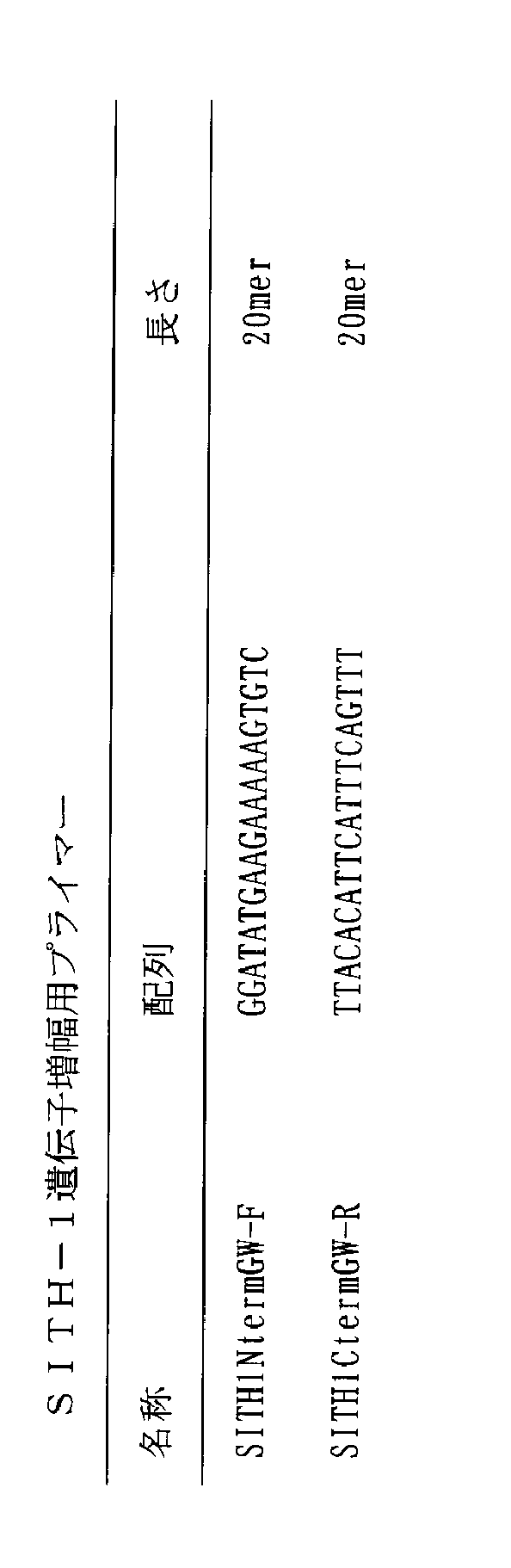
1-2.PCR
SITH-1遺伝子(ORF)(配列番号2)がFLAGタグをつけた発現ベクター(PCT/JP2008/67300)のDNAを鋳型にして、プライマーSITH1NtermGW-FとSITH1CtermGW-Rを用いてSITH-1部位の増幅を行った。PCR反応条件は、20μlの反応液中に鋳型DNAを500ng、10(ExTaq buffer(TaKaRa社)を2μl、2.5mM dNTPを1.6μl、プライマーを各20pmoles、5U/μl ExTaqを0.1μl添加し、GeneAmp PCR System 9600(PERKIN ELMER)を用いて96℃3分を1回、95℃で1分,60℃で1分,72℃で2分を20回、72℃6分を1回行った。その結果、477bpのPCR産物が得られた。 (SEQ ID NO: 10-11)
1-2. PCR
The DNA of the expression vector (PCT / JP2008 / 67300) in which the SITH-1 gene (ORF) (SEQ ID NO: 2) is FLAG-tagged is used as a template, and primers SITH1NtermGW-F and SITH1CtermGW-R are used for the SITH-1 site. Amplification was performed. PCR reaction conditions were as follows: 500 ng of template DNA, 2 μl of 10 (ExTaq buffer (TaKaRa)), 1.6 μl of 2.5 mM dNTP, 20 μmoles of each primer, 0.1 μl of 5 U / μl ExTaq in 20 μl of reaction solution Then, using GeneAmp PCR System 9600 (PERKIN ELMER), 96 ° C for 3 minutes once, 95 ° C for 1 minute, 60 ° C for 1 minute, 72 ° C for 2
PCRによって得られたSITH-1遺伝子を、ベクターpCR8/GW/TOPO(Invitrogen社製)にクローニングした。ライゲーション反応はベクターキット添付の説明書に従った。大腸菌TB1にエレクトロポレーション法を用いてDNAを導入し、常法(Sambrook et al.1989, Molecular Cloning, A laboratory manual, 2nd edition)に従ってプラスミドDNAを抽出した。インサートの存在が確認されたプラスミドに関してM13プライマー(TaKaRa社)と、ABI PRISM蛍光シークエンサー(Model 310 Genetic Analyzer,Perkin Elmer社)を用いて塩基配列を決定し、設計した遺伝子の配列と比較して、変異がないことを確認した。 1-3. Cloning The SITH-1 gene obtained by PCR was cloned into the vector pCR8 / GW / TOPO (Invitrogen). The ligation reaction followed the instructions attached to the vector kit. DNA was introduced into E. coli TB1 using electroporation, and plasmid DNA was extracted according to a conventional method (Sambrook et al. 1989, Molecular Cloning, A laboratory manual, 2nd edition). The base sequence was determined using the M13 primer (TaKaRa) and the ABI PRISM fluorescent sequencer (Model 310 Genetic Analyzer, Perkin Elmer) for the plasmid in which the presence of the insert was confirmed, and compared with the designed gene sequence. It was confirmed that there was no mutation.
BioEase-SITH1/pET104.1を導入した大腸菌BL21(DE3)、又は対照としてpTrc99Aのみを導入した大腸菌BL21を、抗生物質アンピシリン(最終濃度100μg/ml)を含むLB培地50mlに接種し、OD600における吸光度が0.5に達するまで30℃で振とう培養した。その後、1mM IPTGを添加し、さらに30℃で5時間振とう培養した。培養液50mlから遠心にて菌体を回収した。菌体は0.1M HEPES/KOH(pH7.4)3ml中に懸濁後、超音波により破砕した。破砕液を遠心(15,000rpm)し、その上清を大腸菌粗抽出液とした。 1-4. E. coli expression E. coli BL21 (DE3) introduced with BioEase-SITH1 / pET104.1, or E. coli BL21 introduced only with pTrc99A as a control, was inoculated into 50 ml of LB medium containing antibiotic ampicillin (final concentration 100 μg / ml), and OD600 The culture was shaken at 30 ° C. until the absorbance at 0.5 reached 0.5. Thereafter, 1 mM IPTG was added, and further cultured with shaking at 30 ° C. for 5 hours. The cells were collected from 50 ml of the culture solution by centrifugation. The cells were suspended in 3 ml of 0.1 M HEPES / KOH (pH 7.4) and then crushed by ultrasonic waves. The disrupted solution was centrifuged (15,000 rpm), and the supernatant was used as an E. coli crude extract.
精製TM2をNew ELISA plateキット(住友ベークライト)を用いてマイクロプレートに固定化した。固定化方法はキット添付の説明書に従った。 Example 2 Detection of Anti-SITH-1 Antibody in Human Serum by ELISA Purified TM2 was immobilized on a microplate using a New ELISA plate kit (Sumitomo Bakelite). The immobilization method followed the instructions attached to the kit.
ウサギ抗SITH-1抗体(抗血清)の血清中抗体価は、典型的なうつ病(気分障害)患者の血清中抗SITH-1抗体価に比べ、およそ50倍程度高いと推定される。従って市販(健常者)ヒト血清に体積比で1/50量のウサギ抗SITH-1抗体(抗血清)を混ぜると、典型的なうつ病患者血清の擬似検体となると考えられる。よって、上記の抗体希釈率は、うつ病患者血清をおよそ10倍から80倍希釈して用いる場合と同程度とみなすことができる。
さらに、血清の希釈に用いた大腸菌粗抽出液中におけるTM2の効果を確認するために、PBS若しくは上記pTrc99A導入大腸菌粗抽出液に、最終濃度50μg/mlとなるように、精製TM2を加えた溶液を調製し、これを用いてヒト血清を100倍希釈した溶液に、上記と同様、ウサギ抗SITH-1抗体を体積比で1/500、1/1000、1/2000、1/4000、1/8000となるように段階希釈して加え、BioEaseタグ融合SITH-1を固定化したプレートに100μlずつ添加し、1時間室温で反応させた。また対照として、何も結合させていないTM2固定化プレートに上記ブロッキング操作を行った後、上記と同様にPBS溶液や、pTrc99A導入あるいはTM2/pTrc99A導入大腸菌粗抽出液(1mg総可溶性タンパク質/ml)、さらに精製TM2を加えたPBSや、精製TM2を加えたpTrc99A導入大腸菌粗抽出液で、100倍希釈したヒト血清に、段階的に希釈したウサギ抗SITH-1抗体を加えたものを100μlずつ添加し、室温で1時間反応させた。 Next, human serum (Human Serum pool, manufactured by Cosmo-Bio) was prepared using PBS, or 1 mg total soluble protein / ml pTrc99A-introduced Escherichia coli crude extract prepared in Example 1 (1-4), or 1 mg total soluble. In a solution diluted with protein / ml TM2 / pTrc99A-introduced E. coli crude extract 100 times, rabbit anti-SITH-1 antibody was 1/500, 1/1000, 1/2000, 1/4000, 1/8000 in volume ratio. It was added after serial dilution. 100 μl of this diluted human serum (including anti-SITH-1 antibody) was added to a plate on which BioEase tag-fused SITH-1 protein was immobilized using biotin-
The serum antibody titer of rabbit anti-SITH-1 antibody (antiserum) is estimated to be about 50 times higher than the serum anti-SITH-1 antibody titer of typical patients with depression (mood disorder). Therefore, mixing commercially available (healthy person) human serum with a volume ratio of 1/50 rabbit anti-SITH-1 antibody (antiserum) is considered to be a typical specimen of a depression patient serum. Therefore, the above antibody dilution ratio can be regarded as the same level as when the depression patient serum is diluted approximately 10 to 80 times.
Furthermore, in order to confirm the effect of TM2 in the Escherichia coli crude extract used for dilution of serum, a solution obtained by adding purified TM2 to PBS or the pTrc99A-introduced Escherichia coli crude extract to a final concentration of 50 μg / ml. In the same manner as above, the rabbit anti-SITH-1 antibody was added in a volume ratio of 1/500, 1/1000, 1/2000, 1/4000, 1 / 100 μl each was added to a plate on which BioEase tag fusion SITH-1 was immobilized, and the mixture was allowed to react at room temperature for 1 hour. As a control, the blocking operation was performed on a TM2 fixed plate to which nothing was bound, and then PBS solution, pTrc99A-introduced or TM2 / pTrc99A-introduced Escherichia coli crude extract (1 mg total soluble protein / ml) was used. Add 100 μl of PBS with purified TM2 or pTrc99A-introduced Escherichia coli crude extract with purified TM2 to which 100-fold diluted human serum is added with serially diluted rabbit anti-SITH-1 antibody And allowed to react for 1 hour at room temperature.
S/N比=抗SITH-1抗体を各濃度で加えた区の抗SITH-1抗体検出量/抗SITH-1抗体を加えない区の抗SITH-1抗体検出量 Human serum diluted with various diluents as described above was added with serial dilutions of rabbit anti-SITH-1 antibody and reacted with BioEase tag-fused SITH-1 protein immobilized on a carrier. And washed 3 times. Subsequently, a horseradish peroxidase-labeled goat anti-goat anti-SITH-1 antibody conjugated to SITH-1 and human IgG in serum considered to be non-specifically bound in each well were detected. 100 μl each of a mixed solution obtained by diluting rabbit IgG antibody and peroxidase-labeled goat anti-human IgG antibody 5000 times each with TBST was added and allowed to stand at room temperature for 1 hour. Thereafter, the plate was washed 3 times with TBST to detect peroxidase activity. To each well, 100 μl of SuperSignal ELISA Pico Chemiluminescent Substrate (PIERCE) was added to each well and allowed to stand at room temperature for 5 minutes, and then the amount of luminescence was measured with a plate reader Infinite M200 (manufactured by TECAN). In addition, as data values, in each concentration group of the rabbit anti-SITH-1 antibody, the control group sample (the BioEase tag fusion SITH-1 was not fixed to the TM2 plate, but the blocking operation was performed with BSA, and each The amount of luminescence of each group treated with human serum containing a concentration of anti-SITH-1 antibody was also measured, and the value of the control group was the value of the luminescence level of the group to which BioEase tag fusion SITH-1 was immobilized. Deducted from. This was the detected amount of anti-SITH-1 antibody contained in the serum. Furthermore, in order to compare the effect of each serum dilution, the S / N ratio was calculated by the following formula.
S / N ratio = anti-SITH-1 antibody detection amount in the group where anti-SITH-1 antibody was added at each concentration / anti-SITH-1 antibody detection amount in the group where anti-SITH-1 antibody was not added
3-1.TM2プレートとビオチン化SITH-1を使用したヒト血清中抗SITH-1抗体価測定
SITH-1抗体を持つことが判っている患者血清6例(慢性疲労症候群2例、うつ病3例、クローン病1例)と、健常人ではあるが、自己抗体などのバックグラウンドが高いため、蛍光抗体法では判別が難しかった血清6例を用いて血清抗体価測定を行なった。なお、患者血清及び健常人コントロール血清は、東京慈恵会医科大学、大阪大学医学系研究科、又は大阪市立大学医学系研究科の倫理委員会の承認を得て採取、検査を行なった。 Example 3 3. ELISA analysis using patient-derived serum samples 3-1. Measurement of anti-SITH-1 antibody titer in human serum using TM2 plate and biotinylated SITH-1 Six patient sera known to have SITH-1 antibody (2 cases of chronic fatigue syndrome, 3 cases of depression, Crohn's disease Serum antibody titers were measured using 6 sera that were difficult to discriminate by the fluorescent antibody method because of the high background of autoantibodies and the like, although they were healthy individuals (1 example). Patient serum and healthy human control serum were collected and tested with the approval of the Ethics Committee of the Jikei University School of Medicine, Osaka University Graduate School of Medicine, or Osaka City University School of Medicine.
2)大腸菌粗抽出液
BugBuster Protein Extraction Reagent (Novagen社製)を、大腸菌(BL21)ペレット1グラムあたり5ml使用した溶解液を、3,000g、10分間遠心分離した上清(IPTG誘導なし)
3)実施例1のように調製した5mg全タンパク質/mlのTM2発現大腸菌粗抽出液(IPTG誘導有り)
これらをBioEaseタグ融合SITH-1タンパク質を結合したプレートに100μlずつ添加し、1時間室温で静置し、さらにTBSTで3回洗浄した。 1) A solution containing 1% casein in TBS-T (Tris-buffered saline, 0.1% Tween 20);
2) Crude extract of Escherichia coli BugBuster Protein Extraction Reagent (manufactured by Novagen) 5 ml / gram of E. coli (BL21) pellet was centrifuged at 3,000 g for 10 minutes to obtain a supernatant (without IPTG induction)
3) 5 mg total protein / ml TM2 expressing Escherichia coli crude extract prepared as in Example 1 (with IPTG induction)
100 μl of each of these was added to a plate to which the BioEase tag-fused SITH-1 protein was bound, allowed to stand at room temperature for 1 hour, and further washed with TBST three times.
本実施例では、ELISA用プレートとして市販されているQIAGEN社のニッケルプレートに、ヒスチジンタグ(Hisタグ)を付加したSITH-1タンパク質を用いて、ELISA法による抗体測定を行なった。 3-2. Measurement of anti-SITH-1 antibody titer in human serum using nickel plate and His tag fusion SITH-1 protein In this example, a histidine tag (His tag) was added to a QIAGEN nickel plate commercially available as an ELISA plate. Using the added SITH-1 protein, antibody measurement was performed by ELISA.
本実施例では、非常に薄い濃度のSITH-1抗体を検出する場合について、TM2融合SITH-1をビオチン化磁性ビーズへ固定化した系における各種血清希釈液の効果を調査した。 Example 4 Detection of Anti-SITH-1 Antibody in the Presence of Human Serum by ELISA Using Magnetic Beads In this example, TM2 fusion SITH-1 was biotinylated when detecting a very thin concentration of SITH-1 antibody. The effect of various serum dilutions in a system immobilized on magnetic beads was investigated.
SITH1-TM2融合遺伝子構築のために、まず、SITH-1とTM2の両遺伝子をリンカー(5xlinker: GGGGSGGGGSGGGGSGGGGSGGGGS)(配列番号12)を介し融合させるためのプライマーを設計した。 4-1. Primer design To construct the SITH1-TM2 fusion gene, first, a primer for fusing both SITH-1 and TM2 genes via a linker (5xlinker: GGGGSGGGGSGGGGSGGGGSGGGGS) (SEQ ID NO: 12) was designed.
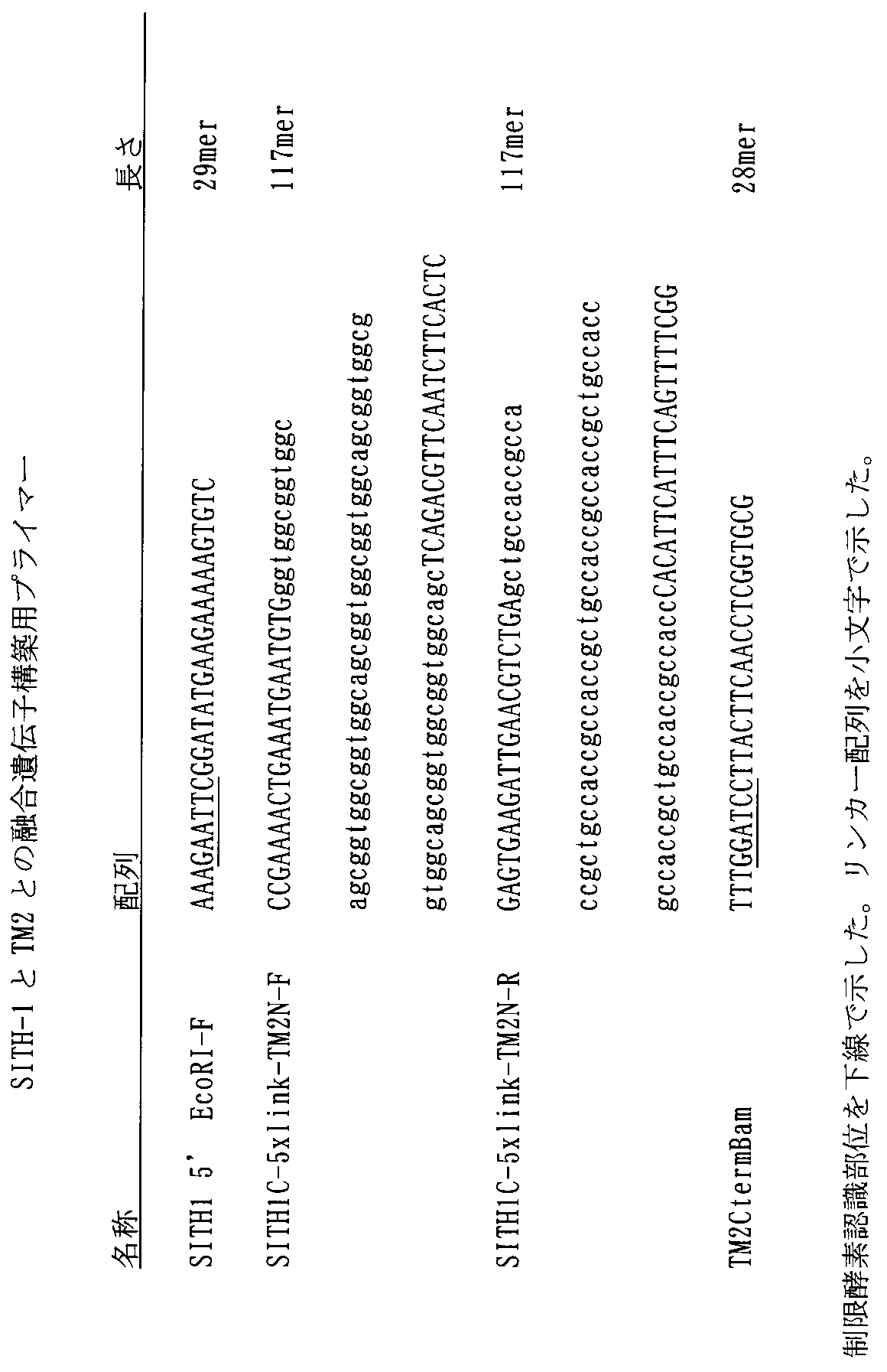
SITH1-TM2融合遺伝子を構築するために、二段階のPCRを行った。 4-2. PCR
To construct the SITH1-TM2 fusion gene, a two-step PCR was performed.
PCRによって得られたSITH1-TM2融合遺伝子をベクターpCR4Blunt TOPO(Invitrogen社製)に、上記実施例1-3と同様にクローニングした。さらに、融合遺伝子が組み込まれたプラスミドをEcoRIとBamHIで二重消化し、融合遺伝子が含まれるDNA断片を回収した。この断片を予めEcoRIとBamHIで消化しておいた大腸菌用の発現ベクターpTrc99A(Pharmacia社製)にクローニングした。以上のように、SITH-1とTM2の融合タンパク質発現用のベクターSITH1-TM2/pTrc99Aを完成させた。発現用のベクターSITH1-TM2/pTrc99A中のSITH1-TM2をコードする塩基配列を配列番号17に、コードされるアミノ酸配列を配列番号18に示す。 4-3. Cloning The SITH1-TM2 fusion gene obtained by PCR was cloned into the vector pCR4Blunt TOPO (Invitrogen) in the same manner as in Example 1-3 above. Furthermore, the plasmid in which the fusion gene was incorporated was double-digested with EcoRI and BamHI, and the DNA fragment containing the fusion gene was recovered. This fragment was cloned into an expression vector pTrc99A (manufactured by Pharmacia) for E. coli that had been digested with EcoRI and BamHI in advance. As described above, a vector SITH1-TM2 / pTrc99A for expressing a fusion protein of SITH-1 and TM2 was completed. The nucleotide sequence encoding SITH1-TM2 in the expression vector SITH1-TM2 / pTrc99A is shown in SEQ ID NO: 17, and the encoded amino acid sequence is shown in SEQ ID NO: 18.
SITH1-TM2/pTrc99Aを導入した大腸菌BL21を、上記実施例1-4.と同様に培養した。培養液50mlから遠心にて菌体を回収した。菌体は0.1M HEPES/KOH(pH7.4)3ml中に懸濁後、超音波により破砕した。破砕液を遠心(15,000rpm)し、その上清を大腸菌粗抽出液とした。TM2融合SITH-1タンパク質の発現をウェスタンブロッティングにより確認した。 4-4. E. coli expression E. coli BL21 into which SITH1-TM2 / pTrc99A was introduced was treated as described in Example 1-4. And cultured in the same manner. The cells were collected from 50 ml of the culture solution by centrifugation. The cells were suspended in 3 ml of 0.1 M HEPES / KOH (pH 7.4) and then crushed by ultrasonic waves. The disrupted solution was centrifuged (15,000 rpm), and the supernatant was used as an E. coli crude extract. The expression of TM2 fusion SITH-1 protein was confirmed by Western blotting.
磁性ビーズのDynabeads M-270 Amine (DynalBiotech社製)1mL(30mgビーズ/mL)に、10mM NHS-Lc-Lc-Biotin (PIERCE社製)1mLを添加した。室温で30分間転倒混和することでアミノ基とNHS活性エステル基を反応させ、ビオチンと磁性ビーズを共有結合させた。その後、磁性ビーズを0.1% BSA/ 0.01% Tween20/ PBS溶液で2回洗浄し、最後にPBS緩衝液に懸濁した。得られた磁性ビーズ(30mgビーズ/mL PBS)をビオチン化磁性ビーズとした。 4-5. Preparation of TM2 Fusion SITH-1 Immobilized
市販のヒト血清(Human Serum pool、Cosmo-Bio社製)に、実施例2のウサギ抗SITH-1抗体を1/50量加えて、SITH-1抗体を含むヒト血清を調製した。一方、SITH-1抗体を加えないヒト血清を対照とした。 4-6. ELISA analysis using magnetic beads Human serum containing SITH-1 antibody by adding 1/50 amount of rabbit anti-SITH-1 antibody of Example 2 to commercially available human serum (Human Serum pool, manufactured by Cosmo-Bio) Was prepared. On the other hand, human serum to which no SITH-1 antibody was added was used as a control.
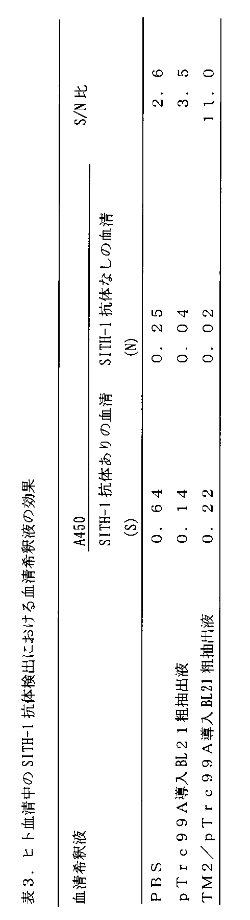
Claims (14)
- 生物学的試料中のHHV-6の潜伏感染中間段階転写物によってコードされる小タンパク質(Small protein encoded by the Intermediate stage Transcript of HHV-6)(SITH-1)の抗体を検出する方法であって、
1) SITH-1タンパク質を準備し;
2) 工程1)で準備したSITH-1タンパク質を担体に結合し;
3) 生物学的試料を、工程2)で作成したSITH-1タンパク質が結合した担体に接触させ、SITH-1タンパク質抗体を検出する
ことを含む、前記方法。 A method for detecting an antibody of Small protein encoded by the intermediate stage Transscript of HHV-6 (SITH-1) encoded by a latently infected intermediate transcript of HHV-6 in a biological sample. ,
1) Prepare SITH-1 protein;
2) Binding the SITH-1 protein prepared in step 1) to a carrier;
3) The method comprising contacting a biological sample with the carrier bound with the SITH-1 protein prepared in step 2), and detecting the SITH-1 protein antibody. - SITH-1タンパク質が以下のグループから選択される、請求項1の方法。
(a) 配列番号1のアミノ酸配列を有するタンパク質;
(b) 配列番号1のアミノ酸配列において、1又は複数のアミノ酸の欠失、置換、挿入及び/又は付加を含むアミノ酸配列を有し、かつ、細胞内カルシウム濃度を上昇させる活性を有するタンパク質;
(c) 配列番号1のアミノ酸配列に対し少なくとも80%の同一性を有するアミノ酸配列を有し、かつ、細胞内カルシウム濃度を上昇させる活性を有するタンパク質;
(d) 配列番号2の塩基配列からなる核酸によりコードされるアミノ酸配列を有するタンパク質;
(e) 配列番号2の塩基配列において1又は複数のヌクレオチドの欠失、置換、挿入及び/又は付加を含む塩基配列からなる核酸によりコードされるアミノ酸配列を有し、かつ、細胞内カルシウム濃度を上昇させる活性を有するタンパク質;
(f) 配列番号2の塩基配列と80%以上の同一性を有する塩基配列からなる核酸によりコードされるアミノ酸配列を有し、かつ、細胞内カルシウム濃度を上昇させる活性を有するタンパク質;及び
(g) 配列番号2の塩基配列に相補的な塩基配列からなる核酸とストリンジェントなハイブリダイゼーション条件下でハイブリダイズする核酸によってコードされ、かつ、細胞内カルシウム濃度を上昇させる活性を有するタンパク質 2. The method of claim 1, wherein the SITH-1 protein is selected from the following group.
(A) a protein having the amino acid sequence of SEQ ID NO: 1;
(B) a protein having an amino acid sequence including deletion, substitution, insertion and / or addition of one or more amino acids in the amino acid sequence of SEQ ID NO: 1 and having an activity of increasing intracellular calcium concentration;
(C) a protein having an amino acid sequence having at least 80% identity to the amino acid sequence of SEQ ID NO: 1 and having an activity of increasing intracellular calcium concentration;
(D) a protein having an amino acid sequence encoded by a nucleic acid consisting of the base sequence of SEQ ID NO: 2;
(E) having an amino acid sequence encoded by a nucleic acid consisting of a base sequence containing a deletion, substitution, insertion and / or addition of one or more nucleotides in the base sequence of SEQ ID NO: 2 and having an intracellular calcium concentration A protein with increasing activity;
(F) a protein having an amino acid sequence encoded by a nucleic acid consisting of a base sequence having 80% or more identity with the base sequence of SEQ ID NO: 2 and having an activity of increasing intracellular calcium concentration; and (g ) A protein encoded by a nucleic acid that hybridizes under stringent hybridization conditions with a nucleic acid comprising a base sequence complementary to the base sequence of SEQ ID NO: 2 and has an activity to increase intracellular calcium concentration - 請求項1の工程3)において、工程2)で作成したSITH-1タンパク質が結合した担体に、
(a) 生物学的試料、及び
(b) 工程1)のSITH-1タンパク質を発現させるのに利用した宿主細胞と同種の細胞より調製した細胞破砕抽出液
を混合して添加する;
請求項1又は2に記載の検出方法 In step 3) of claim 1, the carrier to which the SITH-1 protein prepared in step 2) is bound,
(A) a biological sample, and (b) a cell disruption extract prepared from the same type of cells as the host cell used to express the SITH-1 protein in step 1) is mixed and added;
The detection method according to claim 1 or 2 - 請求項1の工程2)において、SITH-1タンパク質を、ビオチン-ビオチン結合性タンパク質間結合を利用して担体に結合することを特徴とする、請求項1ないし3のいずれか1項に記載の検出方法。 4. The step 2) of claim 1, wherein the SITH-1 protein is bound to a carrier using a biotin-biotin-binding protein-protein bond. Detection method.
- 請求項1の工程3)において、工程2)で作成したSITH-1タンパク質が結合した担体に、
(a) 生物学的試料、並びに
(b-i) 工程1)又は2)のSITH-1タンパク質、ビオチン化SITH-1タンパク質、及び/又はビオチン結合性タンパク質を発現させるのに利用した宿主細胞と同種の細胞より調製した細胞破砕抽出液と、ビオチン結合性タンパク質、又は
(b-ii) 工程1)又は2)のSITH-1タンパク質、ビオチン化SITH-1タンパク質、及び/又はビオチン結合性タンパク質を発現させるのに利用した宿主細胞と同種の細胞に遺伝子工学技術によりビオチン結合性タンパク質を発現させた細胞から調製した細胞破砕抽出液
を混合して添加する;
請求項4に記載の検出方法 In step 3) of claim 1, the carrier to which the SITH-1 protein prepared in step 2) is bound,
(A) a biological sample and (bi) a host cell used to express the SITH-1 protein, biotinylated SITH-1 protein, and / or biotin-binding protein of step 1) or 2) Cell disruption extract prepared from the same type of cells and biotin-binding protein, or (b-ii) SITH-1 protein, biotinylated SITH-1 protein and / or biotin-binding protein of step 1) or 2) Cell disruption extract prepared from cells expressing biotin-binding protein by genetic engineering technology is added to cells of the same type as the host cell used for expression;
The detection method according to claim 4. - 請求項3の工程3(b)又は請求項5の工程3(b-i)において、細胞破砕抽出液として、任意のベクターを含む細胞から抽出した細胞破砕抽出液を添加する、請求項3ないし5のいずれか1項に記載の方法。 In step 3 (b) of claim 3 or step 3 (bi) of claim 5, a cell disruption extract extracted from cells containing an arbitrary vector is added as a cell disruption extract. 6. The method according to any one of 5 above.
- ビオチン結合性タンパク質がタマビジン又はその変異体である、請求項4ないし6のいずれか1項に記載の方法。 The method according to any one of claims 4 to 6, wherein the biotin-binding protein is tamavidin or a variant thereof.
- 生物学的試料が、血液、血清、脳脊髄液、唾液、咽頭拭い液、汗、尿、涙、リンパ液、精液、腹水、及び母乳からなる群から選択される、請求項1ないし6のいずれか7項に記載の方法。 The biological sample is selected from the group consisting of blood, serum, cerebrospinal fluid, saliva, throat swab, sweat, urine, tears, lymph, semen, ascites, and breast milk. 8. The method according to item 7.
- 生物学的試料中のHHV-6の潜伏感染中間段階転写物によってコードされる小タンパク質(Small protein encoded by the Intermediate stage Transcript of HHV-6)(SITH-1)の抗体を検出するための担体であって、前記SITH-1タンパク質を結合した担体。 A carrier for detecting the antibody of Small Protein encoded by the the intermediate stage Transscript of HHV-6 (SITH-1) encoded by a latent intermediate transcript of HHV-6 in a biological sample A carrier to which the SITH-1 protein is bound.
- SITH-1タンパク質と担体とがビオチン-ビオチン結合性タンパク質間結合を利用して結合していることを特徴とする、請求項9に記載の担体。 10. The carrier according to claim 9, wherein the SITH-1 protein and the carrier are bound using a biotin-biotin-binding protein-protein bond.
- 生物学的試料中のHHV-6の潜伏感染中間段階転写物によってコードされる小タンパク質(Small protein encoded by the Intermediate stage Transcript of HHV-6)(SITH-1)の抗体を検出するためのキットであって、
A) 前記SITH-1タンパク質が結合している担体;並びに、
B) A)のSITH-1タンパク質を発現させるのに利用した宿主細胞と同種の細胞より調製した細胞破砕抽出液を含む、生物学的試料を希釈するための剤
を含むキット。 A kit for detecting an antibody of Small protein encoded by the intermediate stage Transscript of HHV-6 (SITH-1) encoded in a latent sample of latent infection of HHV-6 in a biological sample There,
A) a carrier to which the SITH-1 protein is bound; and
B) A kit comprising an agent for diluting a biological sample, comprising a cell disruption extract prepared from cells of the same type as the host cell used to express the SITH-1 protein of A). - A)の担体が、SITH-1タンパク質がビオチン-ビオチン結合性タンパク質間結合を利用して結合している担体であり、そして、
B)の剤が、
i) A)のSITH-1タンパク質、ビオチン化SITH-1タンパク質、及び/又はビオチン結合性タンパク質を発現させるのに利用した宿主細胞と同種の細胞より調製した細胞破砕抽出液と、ビオチン結合性タンパク質、又は
ii) A)のSITH-1タンパク質、ビオチン化SITH-1タンパク質、及び/又はビオチン結合性タンパク質を発現させるのに利用した宿主細胞と同種の細胞に遺伝子工学技術によりビオチン結合性タンパク質を発現させた細胞から調製した細胞破砕抽出液
を含む、生物学的試料を希釈するための剤である、
請求項11に記載のキット。 The carrier of A) is a carrier in which the SITH-1 protein is bound using a biotin-biotin-binding protein-protein bond, and
The agent of B)
i) A cell disruption extract prepared from cells of the same type as the host cell used to express the SITH-1 protein, biotinylated SITH-1 protein, and / or biotin-binding protein of A), and a biotin-binding protein Or ii) A biotin-binding protein is expressed by genetic engineering technology in the same type of host cell used to express the SITH-1 protein, biotinylated SITH-1 protein, and / or biotin-binding protein of A) An agent for diluting a biological sample, including a cell disruption extract prepared from the treated cells,
The kit according to claim 11. - 生物学的試料中のHHV-6の潜伏感染中間段階転写物によってコードされる小タンパク質(Small protein encoded by the Intermediate stage Transcript of HHV-6)(SITH-1)の抗体を検出するためのキットであって、
A) 前記SITH-1タンパク質;
B) A)のSITH-1タンパク質を固定化するための担体;並びに
C) A)のSITH-1タンパク質を発現させるのに利用した宿主細胞と同種の細胞より調製した細胞破砕抽出液を含む、生物学的試料を希釈するための剤
を含むキット。 A kit for detecting an antibody of Small protein encoded by the intermediate stage Transscript of HHV-6 (SITH-1) encoded in a latent sample of latent infection of HHV-6 in a biological sample There,
A) the SITH-1 protein;
B) a carrier for immobilizing the SITH-1 protein of A); and C) a cell disruption extract prepared from cells of the same type as the host cell used to express the SITH-1 protein of A). A kit containing an agent for diluting a biological sample. - A)のSITH-1タンパク質がビオチン化されており、
B)の担体が、ビオチン結合性タンパク質と直接的又は間接的に結合しており、
C)の剤が、
i) A)又はB)のSITH-1タンパク質、ビオチン化SITH-1タンパク質、及び/又はビオチン結合性タンパク質を発現させるのに利用した宿主細胞と同種の細胞より調製した細胞破砕抽出液と、ビオチン結合性タンパク質、又は
ii) A)又はB)のSITH-1タンパク質、ビオチン化SITH-1タンパク質、及び/又はビオチン結合性タンパク質を発現させるのに利用した宿主細胞と同種の細胞に遺伝子工学技術によりビオチン結合性タンパク質を発現させた細胞から調製した細胞破砕抽出液
を含む、生物学的試料を希釈するための剤である、
請求項13に記載のキット。 A) SITH-1 protein is biotinylated,
The carrier of B) is bound directly or indirectly to the biotin-binding protein,
C)
i) A cell disruption extract prepared from cells of the same type as the host cell used to express the SITH-1 protein, biotinylated SITH-1 protein, and / or biotin-binding protein of A) or B), and biotin Genetic engineering techniques to bind proteins, or ii) cells of the same type as the host cell used to express the SITH-1 protein, biotinylated SITH-1 protein, and / or biotin binding protein of A) or B) An agent for diluting a biological sample containing a cell disruption extract prepared from cells expressing a biotin-binding protein.
The kit according to claim 13.
Priority Applications (10)
| Application Number | Priority Date | Filing Date | Title |
|---|---|---|---|
| EP10758800A EP2416159A4 (en) | 2009-03-31 | 2010-03-31 | Method for detecting antibody against sith-1 in biological sample |
| US13/257,754 US9139617B2 (en) | 2009-03-31 | 2010-03-31 | Method for detecting antibody against SITH-1 in biological sample |
| JP2011507263A JPWO2010114029A1 (en) | 2009-03-31 | 2010-03-31 | Method for detecting antibodies of SITH-1 in a biological sample |
| BRPI1015458A BRPI1015458A2 (en) | 2009-03-31 | 2010-03-31 | method for detecting sith-1 antibody in biological sample |
| RU2011143788/15A RU2011143788A (en) | 2009-03-31 | 2010-03-31 | METHOD FOR DETECTING ANTI-SITH-1 ANTIBODIES IN A BIOLOGICAL SAMPLE |
| CN2010800239031A CN102449483A (en) | 2009-03-31 | 2010-03-31 | Method for detecting antibody against sith-1 in biological sample |
| AU2010232303A AU2010232303A1 (en) | 2009-03-31 | 2010-03-31 | Method for detecting antibody against SITH-1 in biological sample |
| SG2011070216A SG174938A1 (en) | 2009-03-31 | 2010-03-31 | Method for detecting antibody against sith-1 in biological sample |
| CA 2756006 CA2756006A1 (en) | 2009-03-31 | 2010-03-31 | Method for detecting antibody against sith-1 in biological sample |
| IL215322A IL215322A0 (en) | 2009-03-31 | 2011-09-22 | Method for detecting antibody against sith-1 in biological sample |
Applications Claiming Priority (2)
| Application Number | Priority Date | Filing Date | Title |
|---|---|---|---|
| JP2009087816 | 2009-03-31 | ||
| JP2009-087816 | 2009-03-31 |
Publications (1)
| Publication Number | Publication Date |
|---|---|
| WO2010114029A1 true WO2010114029A1 (en) | 2010-10-07 |
Family
ID=42828318
Family Applications (1)
| Application Number | Title | Priority Date | Filing Date |
|---|---|---|---|
| PCT/JP2010/055884 WO2010114029A1 (en) | 2009-03-31 | 2010-03-31 | Method for detecting antibody against sith-1 in biological sample |
Country Status (13)
| Country | Link |
|---|---|
| US (1) | US9139617B2 (en) |
| EP (1) | EP2416159A4 (en) |
| JP (1) | JPWO2010114029A1 (en) |
| KR (1) | KR20110137379A (en) |
| CN (1) | CN102449483A (en) |
| AU (1) | AU2010232303A1 (en) |
| BR (1) | BRPI1015458A2 (en) |
| CA (1) | CA2756006A1 (en) |
| IL (1) | IL215322A0 (en) |
| RU (1) | RU2011143788A (en) |
| SG (1) | SG174938A1 (en) |
| TW (1) | TW201040529A (en) |
| WO (1) | WO2010114029A1 (en) |
Cited By (5)
| Publication number | Priority date | Publication date | Assignee | Title |
|---|---|---|---|---|
| EP2416158A1 (en) * | 2009-03-31 | 2012-02-08 | Japan Tobacco, Inc. | Method for detecting substance in biological sample |
| JP2015500502A (en) * | 2011-12-13 | 2015-01-05 | バクスター、インターナショナル、インコーポレイテッド | Autoantibody assay with high sensitivity under low conductivity conditions |
| WO2015133439A1 (en) * | 2014-03-07 | 2015-09-11 | 日本全薬工業株式会社 | Pullulan gel, method for producing same, and use of same |
| WO2017115878A1 (en) * | 2015-12-28 | 2017-07-06 | 日本たばこ産業株式会社 | Method for diagnosing, treating or preventing mood disorders |
| US10487118B2 (en) | 2007-09-27 | 2019-11-26 | Virus Ikagaku Kenkyusho Inc. | Factor involved in latent infection with herpes virus, and use thereof |
Families Citing this family (2)
| Publication number | Priority date | Publication date | Assignee | Title |
|---|---|---|---|---|
| CA2753873A1 (en) * | 2009-03-02 | 2010-09-10 | Japan Tobacco Inc. | Method for detecting substance in biological sample |
| EP3163301B1 (en) * | 2014-06-27 | 2019-08-07 | Japan Tobacco, Inc. | Method for treating, or preventing mood disorder |
Citations (10)
| Publication number | Priority date | Publication date | Assignee | Title |
|---|---|---|---|---|
| JPS5999257A (en) | 1982-11-29 | 1984-06-07 | Green Cross Corp:The | Aqueous solvent of reagent for detecting antibody |
| JPH02233697A (en) * | 1989-01-23 | 1990-09-17 | Boehringer Mannheim Gmbh | Measurement of hiv antibody and biotinized polypeptide |
| JPH0843392A (en) | 1994-07-29 | 1996-02-16 | Fujirebio Inc | Nonspecific reactive absorbent for antibody sensing reagent |
| WO2002072817A1 (en) | 2001-03-12 | 2002-09-19 | Japan Tobacco Inc. | Novel protein, gene encoding the same and method of using the same |
| JP2004301646A (en) | 2003-03-31 | 2004-10-28 | Sysmex Corp | Nonspecific reaction absorbent for immunity inspection |
| JP2008067300A (en) | 2006-09-11 | 2008-03-21 | Nippon Sheet Glass Co Ltd | Rear window glass for vehicles |
| JP2008261841A (en) * | 2007-03-16 | 2008-10-30 | Sysmex Corp | Hcv antibody measuring reagent kit and hcv antibody measuring method |
| WO2009028625A1 (en) * | 2007-08-28 | 2009-03-05 | Japan Tobacco Inc. | Method of binding protein to support using tamavidin |
| JP2009064302A (en) | 2007-09-07 | 2009-03-26 | Koji Kobayashi | Automatic vending machine with full-face advertisement equipped with donation function |
| WO2009041501A1 (en) * | 2007-09-27 | 2009-04-02 | Virus Ikagaku Kenkyusho Inc. | Factor involved in latent infection with herpesvirus, and use thereof |
Family Cites Families (10)
| Publication number | Priority date | Publication date | Assignee | Title |
|---|---|---|---|---|
| EP1572946B1 (en) * | 2002-09-06 | 2012-03-14 | Amgen, Inc. | Therapeutic human anti-il-1r1 monoclonal antibody |
| CN101351559A (en) * | 2005-11-09 | 2009-01-21 | 普里梅拉生物系统有限公司 | Multiplexed quantitative detection of pathogens |
| PL2007910T3 (en) * | 2006-03-31 | 2017-04-28 | Mobidiag Oy | Method and microarray for detecting herpesviruses |
| US20080176340A1 (en) * | 2006-11-01 | 2008-07-24 | Beckman Coulter, Inc. | Binding surfaces for affinity assays |
| JP4975600B2 (en) * | 2007-03-16 | 2012-07-11 | シスメックス株式会社 | Reagent kit for HCV antibody measurement and HCV antibody measurement method |
| EP1978365B1 (en) | 2007-03-16 | 2010-08-25 | Sysmex Corporation | Reagent kit and method for measuring HCV antibody |
| JP2008261641A (en) | 2007-04-10 | 2008-10-30 | Matsushita Electric Ind Co Ltd | Route guidance device |
| US7972804B2 (en) * | 2007-07-03 | 2011-07-05 | One Lambda | Methods of detecting antibodies specific for denatured HLA antigens |
| CA2753873A1 (en) * | 2009-03-02 | 2010-09-10 | Japan Tobacco Inc. | Method for detecting substance in biological sample |
| US9034590B2 (en) * | 2009-03-31 | 2015-05-19 | Japan Tobacco Inc. | Method for detecting substance in biological sample |
-
2010
- 2010-03-31 RU RU2011143788/15A patent/RU2011143788A/en unknown
- 2010-03-31 WO PCT/JP2010/055884 patent/WO2010114029A1/en active Application Filing
- 2010-03-31 SG SG2011070216A patent/SG174938A1/en unknown
- 2010-03-31 CA CA 2756006 patent/CA2756006A1/en not_active Abandoned
- 2010-03-31 KR KR20117025224A patent/KR20110137379A/en not_active Application Discontinuation
- 2010-03-31 JP JP2011507263A patent/JPWO2010114029A1/en active Pending
- 2010-03-31 CN CN2010800239031A patent/CN102449483A/en active Pending
- 2010-03-31 TW TW99109999A patent/TW201040529A/en unknown
- 2010-03-31 BR BRPI1015458A patent/BRPI1015458A2/en not_active Application Discontinuation
- 2010-03-31 US US13/257,754 patent/US9139617B2/en active Active
- 2010-03-31 EP EP10758800A patent/EP2416159A4/en not_active Withdrawn
- 2010-03-31 AU AU2010232303A patent/AU2010232303A1/en not_active Abandoned
-
2011
- 2011-09-22 IL IL215322A patent/IL215322A0/en unknown
Patent Citations (10)
| Publication number | Priority date | Publication date | Assignee | Title |
|---|---|---|---|---|
| JPS5999257A (en) | 1982-11-29 | 1984-06-07 | Green Cross Corp:The | Aqueous solvent of reagent for detecting antibody |
| JPH02233697A (en) * | 1989-01-23 | 1990-09-17 | Boehringer Mannheim Gmbh | Measurement of hiv antibody and biotinized polypeptide |
| JPH0843392A (en) | 1994-07-29 | 1996-02-16 | Fujirebio Inc | Nonspecific reactive absorbent for antibody sensing reagent |
| WO2002072817A1 (en) | 2001-03-12 | 2002-09-19 | Japan Tobacco Inc. | Novel protein, gene encoding the same and method of using the same |
| JP2004301646A (en) | 2003-03-31 | 2004-10-28 | Sysmex Corp | Nonspecific reaction absorbent for immunity inspection |
| JP2008067300A (en) | 2006-09-11 | 2008-03-21 | Nippon Sheet Glass Co Ltd | Rear window glass for vehicles |
| JP2008261841A (en) * | 2007-03-16 | 2008-10-30 | Sysmex Corp | Hcv antibody measuring reagent kit and hcv antibody measuring method |
| WO2009028625A1 (en) * | 2007-08-28 | 2009-03-05 | Japan Tobacco Inc. | Method of binding protein to support using tamavidin |
| JP2009064302A (en) | 2007-09-07 | 2009-03-26 | Koji Kobayashi | Automatic vending machine with full-face advertisement equipped with donation function |
| WO2009041501A1 (en) * | 2007-09-27 | 2009-04-02 | Virus Ikagaku Kenkyusho Inc. | Factor involved in latent infection with herpesvirus, and use thereof |
Non-Patent Citations (29)
| Title |
|---|
| "Molecular Cloning, A Laboratory Manual", 2001, COLD SPRING HARBOR PRESS |
| ALTSCHUL, NUCL. ACIDS. RES., vol. 25, 1997, pages 3389 - 3402 |
| BIOCHEM. J., vol. 363, 2002, pages 609 - 617 |
| BIOCHEM. J., vol. 405, 2007, pages 397 - 405 |
| DEVEREUX ET AL., NUCL. ACIDS RES., vol. 12, 1984, pages 387 |
| DOMINGUEZ ET AL.: "Human Herpesvirus 6B Genome Sequence: Coding Content and Comparison with Human Herpesvirus 6A", JOURNAL OF VIROLOGY, vol. 73, no. 10, 1999, pages 8040 - 8052, XP002403841 * |
| HARLOW ET AL.: "Antibodies: A laboratory manual", 1988, COLD SPRING HARBOR LABORATORY |
| HENIKOFF, S., HENIKOFF, J. G., PROC. NATL. ACAD. SCI. USA, vol. 89, 1992, pages 10915 - 10919 |
| HIRSCH ET AL., BIOCHEM, vol. 308, 2002, pages 343 - 357 |
| HOFMANN ET AL., PROC NATL ACAD SCI USA, vol. 77, 1980, pages 4666 - 4668 |
| IACOBELLI ET AL., BREAST CANCER RESEARCH AND TREATMENT, vol. 11, 1988, pages 19 - 30 |
| IWASAKI: "Monoclonal Antibody: Hybridoma and ELISA", 1991, KODANSHA |
| J. BIOL. CHEM., vol. 280, 2005, pages 13250 - 13255 |
| KOBAYASHI ET AL.: "Human Herpesviruses (HHV)-6 Senpuku Kansen Chukan Jotai Tokuiteki Tanpaku ni yoru Kibun Shogai no Hassho Kijo", DAI 56 KAI THE JAPANESE SOCIETY OF VIROLOGY GAKUJUTSU SHUKAI PROGRAM SHOROKUSHU, 1 October 2008 (2008-10-01), XP009158336 * |
| KONDO ET AL.: "Detection of Gene Cluster That Is Dispensable for Human Herpesvirus 6 Replication abd Latency", JOURNAL OF VIROLOGY, vol. 77, no. 19, 2003, pages 10719 - 10724, XP002903424 * |
| KONDO ET AL.: "Identification of Human Herpesvirus 6 Latency-Associated Transcripts", JOURNAL OF VIROLOGY, vol. 76, no. 8, 2002, pages 4145 - 4151, XP003009785 * |
| KONDO K ET AL.: "Recognition of a Novel Stage of Beta-Herpesvirus Latency in Human Herpesvirus 6.", J VIROL., vol. 77, 2003, pages 2258 - 2264 |
| KONDO KAZUHIRO: "Herpes viruses kansen to hiroh (Infection with herpes viruses and fatigue", VIRUS, vol. 55, no. 1, 2005, pages 9 - 18 |
| KONDO. K ET AL.: "Association of human herpesvirus 6 infection of the central nervous system with recurrence of febrile convulsions", J INFECT DIS, vol. 167, 1993, pages 1197 - 1200 |
| KONDO. K ET AL.: "Identification of human herpesvirus 6 latency-associated transcripts.", J VIROL., vol. 76, 2002, pages 4145 - 4151 |
| KONDO. K ET AL.: "Latent human herpesvirus 6 infection of human monocytes /macrophages", J GEN VIROL, vol. 72, 1991, pages 1401 - 1408 |
| KONDO: "Human Herpesvirus Latency and Fatigue", VIRUS, vol. 55, no. 1, 2005, pages 9 - 18, XP008123498 * |
| KYTE ET AL., J. MOL. BIOL., vol. 157, 1982, pages 105 - 131 |
| NEEDLEMAN, S. B., WUNSCH, C. D., J. MOL. BIOL., vol. 48, 1970, pages 443 - 453 |
| SAMBROOK ET AL.: "Molecular Cloning, A laboratory manual", 1989 |
| SCHWARZ ET AL., J. BIOL. CHEM., vol. 263, 1988, pages 9640 - 9645 |
| See also references of EP2416159A4 |
| TAKAKURA ET AL., FEBS J, vol. 276, 2009, pages 1383 - 1397 |
| TAKAKURA ET AL., FEBS J., vol. 276, 2009, pages 1383 - 1397 |
Cited By (14)
| Publication number | Priority date | Publication date | Assignee | Title |
|---|---|---|---|---|
| US10487118B2 (en) | 2007-09-27 | 2019-11-26 | Virus Ikagaku Kenkyusho Inc. | Factor involved in latent infection with herpes virus, and use thereof |
| EP2416158A4 (en) * | 2009-03-31 | 2012-05-30 | Japan Tobacco Inc | Method for detecting substance in biological sample |
| JPWO2010114031A1 (en) * | 2009-03-31 | 2012-10-11 | 日本たばこ産業株式会社 | Method for detecting a substance in a biological sample |
| JP5628793B2 (en) * | 2009-03-31 | 2014-11-19 | 日本たばこ産業株式会社 | Method for detecting a substance in a biological sample |
| US9034590B2 (en) | 2009-03-31 | 2015-05-19 | Japan Tobacco Inc. | Method for detecting substance in biological sample |
| EP2416158A1 (en) * | 2009-03-31 | 2012-02-08 | Japan Tobacco, Inc. | Method for detecting substance in biological sample |
| JP2015500502A (en) * | 2011-12-13 | 2015-01-05 | バクスター、インターナショナル、インコーポレイテッド | Autoantibody assay with high sensitivity under low conductivity conditions |
| WO2015133439A1 (en) * | 2014-03-07 | 2015-09-11 | 日本全薬工業株式会社 | Pullulan gel, method for producing same, and use of same |
| JPWO2015133439A1 (en) * | 2014-03-07 | 2017-04-06 | 日本全薬工業株式会社 | Pull-langer and its production method and use |
| WO2017115878A1 (en) * | 2015-12-28 | 2017-07-06 | 日本たばこ産業株式会社 | Method for diagnosing, treating or preventing mood disorders |
| CN108431046A (en) * | 2015-12-28 | 2018-08-21 | 日本烟草产业株式会社 | Diagnosis, the method for treating or preventing mood disorder |
| JPWO2017115878A1 (en) * | 2015-12-28 | 2018-12-13 | 日本たばこ産業株式会社 | Method for diagnosing, treating or preventing mood disorders |
| EP3398969A4 (en) * | 2015-12-28 | 2019-07-31 | Japan Tobacco, Inc. | Method for diagnosing, treating or preventing mood disorders |
| US10539578B2 (en) | 2015-12-28 | 2020-01-21 | Japan Tobacco Inc. | Method for diagnosing, treating, or preventing mood disorders |
Also Published As
| Publication number | Publication date |
|---|---|
| SG174938A1 (en) | 2011-11-28 |
| AU2010232303A1 (en) | 2011-11-10 |
| RU2011143788A (en) | 2013-05-10 |
| CN102449483A (en) | 2012-05-09 |
| EP2416159A4 (en) | 2012-05-30 |
| BRPI1015458A2 (en) | 2016-04-19 |
| KR20110137379A (en) | 2011-12-22 |
| TW201040529A (en) | 2010-11-16 |
| IL215322A0 (en) | 2011-12-29 |
| CA2756006A1 (en) | 2010-10-07 |
| JPWO2010114029A1 (en) | 2012-10-11 |
| US20120107842A1 (en) | 2012-05-03 |
| US9139617B2 (en) | 2015-09-22 |
| EP2416159A1 (en) | 2012-02-08 |
Similar Documents
| Publication | Publication Date | Title |
|---|---|---|
| JP5628793B2 (en) | Method for detecting a substance in a biological sample | |
| WO2010114029A1 (en) | Method for detecting antibody against sith-1 in biological sample | |
| JP5647599B2 (en) | Method for detecting a substance in a biological sample | |
| CN111610327B (en) | Novel coronavirus detection kit and preparation method thereof | |
| JPWO2009123347A1 (en) | Virus quantification method | |
| CN113447658B (en) | Kit for detecting anti-peroxiredoxin-1-IgG antibody | |
| JP2023011764A (en) | Method of immobilizing lectin | |
| EP3258265B1 (en) | Method for assessing risk of human cytomegalovirus active infection in body and related kit | |
| JPWO2013018836A1 (en) | Method for suppressing non-specific binding in a step of detecting a substance in a biological sample, and agent for use in the method | |
| KR101419791B1 (en) | A Composition and a Kit for Detecting Specific Antibodies for Orientia tsutsugamushi | |
| US20220196672A1 (en) | Rocky Mountain Spotted Fever Detection and Treatment | |
| Blatch et al. | Bassem SS Guirgis, Cláudia Sá e Cunha, Inês Gomes, Miguel Cavadas, Isabel Silva, Gonçalo Doria, Gregory |
Legal Events
| Date | Code | Title | Description |
|---|---|---|---|
| WWE | Wipo information: entry into national phase |
Ref document number: 201080023903.1 Country of ref document: CN |
|
| 121 | Ep: the epo has been informed by wipo that ep was designated in this application |
Ref document number: 10758800 Country of ref document: EP Kind code of ref document: A1 |
|
| WWE | Wipo information: entry into national phase |
Ref document number: 2011507263 Country of ref document: JP |
|
| WWE | Wipo information: entry into national phase |
Ref document number: 2756006 Country of ref document: CA |
|
| WWE | Wipo information: entry into national phase |
Ref document number: 13257754 Country of ref document: US |
|
| NENP | Non-entry into the national phase |
Ref country code: DE |
|
| WWE | Wipo information: entry into national phase |
Ref document number: 2010758800 Country of ref document: EP |
|
| ENP | Entry into the national phase |
Ref document number: 20117025224 Country of ref document: KR Kind code of ref document: A |
|
| WWE | Wipo information: entry into national phase |
Ref document number: 7826/CHENP/2011 Country of ref document: IN |
|
| ENP | Entry into the national phase |
Ref document number: 2011143788 Country of ref document: RU Kind code of ref document: A |
|
| ENP | Entry into the national phase |
Ref document number: 2010232303 Country of ref document: AU Date of ref document: 20100331 Kind code of ref document: A |
|
| REG | Reference to national code |
Ref country code: BR Ref legal event code: B01A Ref document number: PI1015458 Country of ref document: BR |
|
| ENP | Entry into the national phase |
Ref document number: PI1015458 Country of ref document: BR Kind code of ref document: A2 Effective date: 20110930 |